Folate Receptor alpha antibody


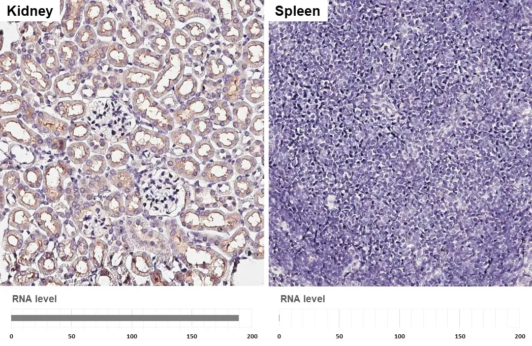
Corresponding RNA levels (RPKM) in the tissues are based on NCBI database.
Folate Receptor alpha antibody detects Folate Receptor alpha protein by immunohistochemical analysis.Sample: Paraffin-embedded mouse tissues.Folate Receptor alpha stained by Folate Receptor alpha antibody (GTX134660) diluted at 1:4000.Antigen Retrieval: Tris-EDTA buffer, pH 9.0, 15 min
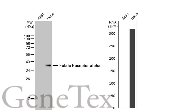
Various whole cell extracts (30 μg) were separated by 12% SDS-PAGE, and the membrane was blotted with Folate Receptor alpha antibody (GTX134660) diluted at 1:500. The HRP-conjugated anti-rabbit IgG antibody (GTX213110-01) was used to detect the primary antibody. Corresponding RNA expression data for the same cell lines are based on Human Protein Atlas program.
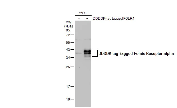
Non-transfected (–) and transfected (+) 293T whole cell extracts (30 μg) were separated by 12% SDS-PAGE, and the membrane was blotted with Folate Receptor alpha antibody (GTX134660) diluted at 1:1000. The HRP-conjugated anti-rabbit IgG antibody (GTX213110-01) was used to detect the primary antibody.
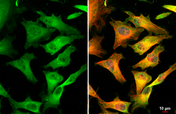
Folate Receptor alpha antibody detects Folate Receptor alpha protein at cytoplasm by immunofluorescent analysis.Sample: HeLa cells were fixed in 4% paraformaldehyde at RT for 15 min.Green: Folate Receptor alpha stained by Folate Receptor alpha antibody (GTX134660) diluted at 1:500.Red: alpha Tubulin, a cytoskeleton marker, stained by alpha Tubulin antibody [GT114] (GTX628802) diluted at 1:1000.Blue: Fluoroshield with DAPI (GTX30920).Scale bar= 10μm.
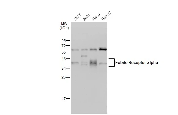
Various whole cell extracts (30 μg) were separated by 12% SDS-PAGE, and the membrane was blotted with Folate Receptor alpha antibody (GTX134660) diluted at 1:500. The HRP-conjugated anti-rabbit IgG antibody (GTX213110-01) was used to detect the primary antibody, and the signal was developed with Trident ECL plus-Enhanced.
-
HostRabbit
-
ClonalityPolyclonal
-
IsotypeIgG
-
ApplicationsWB ICC/IF IHC-P
-
ReactivityHuman, Mouse