GABARAP antibody


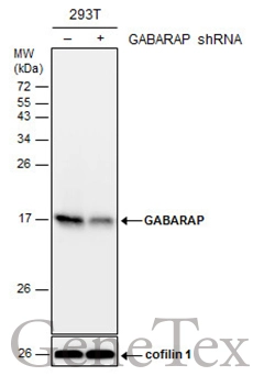
Non-transfected (–) and transfected (+) 293T whole cell extracts (30 μg) were separated by 15% SDS-PAGE, and the membrane was blotted with GABARAP antibody (GTX129710) diluted at 1:1000.
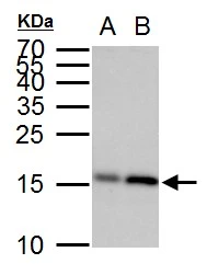
GABARAP antibody detects GABARAP protein by western blot analysis.
A. 30 μg 293T whole cell lysate/extract
B. 30 whole cell lysate/extract of human GABARAP -transfected 293T cells
15 % SDS-PAGE
GABARAP antibody (GTX129710) dilution: 1:10000
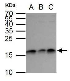
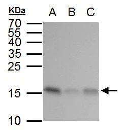
GABARAP antibody detects GABARAP protein by western blot analysis.
A. 30 μg Jurkat whole cell lysate/extract
B. 30 μg Raji whole cell lysate/extract
C. 30 μg NCI-H929 whole cell lysate/extract
15 % SDS-PAGE
GABARAP antibody (GTX129710) dilution: 1:2000
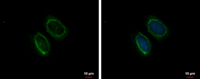
GABARAP antibody detects GABARAP protein at cytoplasm by immunofluorescent analysis.
Sample: U2OS cells were fixed in ice-cold MeOH for 5 min.
Green: GABARAP protein stained by GABARAP antibody (GTX129710) diluted at 1:500.
Blue: Hoechst 33342 staining.
GABARAP antibody detects GABARAP protein by western blot analysis.
A. 30 μg 293T whole cell lysate/extract
B. 30 μg A431 whole cell lysate/extract
C. 30 μg HeLa whole cell lysate/extract
15 % SDS-PAGE
GABARAP antibody (GTX129710) dilution: 1:2000
-
HostRabbit
-
ClonalityPolyclonal
-
IsotypeIgG
-
ApplicationsWB ICC/IF
-
ReactivityHuman