GLI3 antibody

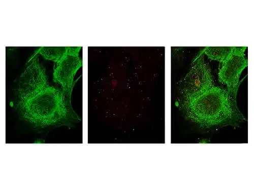
ICC/IF analysis of MCF-7 cells using GTX26050 GLI3 antibody.
Red : Primary antibody
Green : alpha Tubulin
Dilution : 5 μg/mL
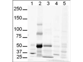
WB analysis of various samples using GTX26050 GLI3 antibody.
Lane 1 : Human brain tissue lysate
Lane 2 : Human lung tissue lysate
Lane 3 : Human spleen tissue lysate
Lane 4 : Mouse brain tissue lysate
Lane 5 : Mouse lung tissue lysate
Loading : 20 μg
Dilution : 1:500

WB analysis of non-transfected and transfected 293T whole cell lysate using GTX26050 GLI3 antibody.
Loading : 35 μg
Dilution : 1:400
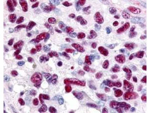
IHC-P analysis of human glioblastoma tissue using GTX26050 GLI3 antibody.
Dilution : 0.625 μg/mL
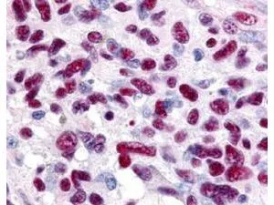
Immunohistochemistry of Rabbit anti-Gli-3 antibody (GTX26050). This image tissue: human glioblastoma. Specific staining was also noted in tissue from adrenal, brain, glioblastoma, colon, heart, kidney, lung, liver, skeletal muscle, ovary, pancreas, placenta, skin, spleen, stomach, testes, thymus, thyroid, tonsil and uterus. Fixation: formalin fixed paraffin embedded. Antigen retrieval: not required. Primary antibody: Gli-3 antibody at 0.625 μg/ml for 1 h at RT. Secondary antibody: Peroxidase rabbit secondary antibody at 1:10,000 for 45 min at RT. Localization: Gli-3 is nuclear and smooth muscle. Staining: Gli-3 as precipitated red signal with hematoxylin purple nuclear counterstain.
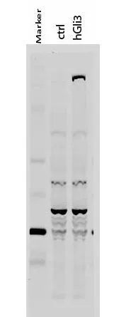
Western Blot of Rabbit anti-Gli-3 antibody. Lane 1: 50 kDa molecular weight marker. Lane 2: 293T cells transfected with CrkL-Flag. Lane 3: 293T cells transfected with human Gli-3. Load: 35 μg per lane. Primary antibody: Gli-3 antibody at 1:400 for overnight at 4ºC. Secondary antibody: IRDye800™ rabbit secondary antibody at 1:10,000 for 45 min at RT. Block: 5% BLOTTO overnight at 4ºC. Predicted/Observed size: 170-190 kDa for hGli-3. Other band(s): Non specific background ~60kDa.
-
HostRabbit
-
ClonalityPolyclonal
-
IsotypeIgG
-
ApplicationsWB ICC/IF IHC-P ELISA Multiplexing
-
ReactivityHuman, Fish