GOT2 antibody

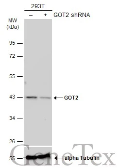
Non-transfected (–) and transfected (+) 293T whole cell extracts (30 μg) were separated by 10% SDS-PAGE, and the membrane was blotted with GOT2 antibody (GTX101930) diluted at 1:20000. The HRP-conjugated anti-rabbit IgG antibody (GTX213110-01) was used to detect the primary antibody.
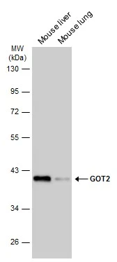
Various tissue extracts (50 μg) were separated by 10% SDS-PAGE, and the membrane was blotted with GOT2 antibody (GTX101930) diluted at 1:10000. The HRP-conjugated anti-rabbit IgG antibody (GTX213110-01) was used to detect the primary antibody, and the signal was developed with Trident ECL plus-Enhanced.
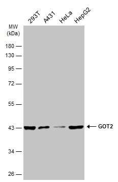
Various whole cell extracts (30 μg) were separated by 10% SDS-PAGE, and the membrane was blotted with GOT2 antibody (GTX101930) diluted at 1:10000. The HRP-conjugated anti-rabbit IgG antibody (GTX213110-01) was used to detect the primary antibody.
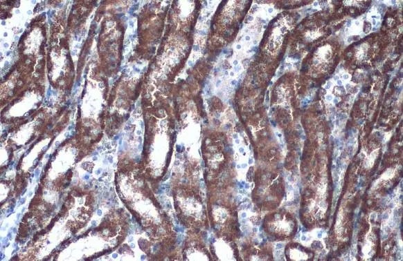
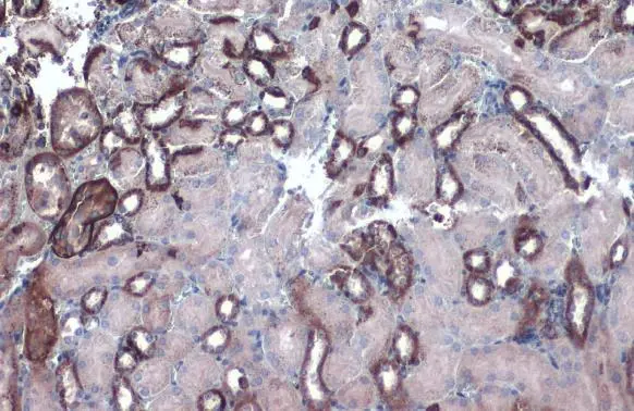
GOT2 antibody detects GOT2 protein at mitochondria by immunohistochemical analysis.Sample: Paraffin-embedded rat kidney.GOT2 stained by GOT2 antibody (GTX101930) diluted at 1:1000.Antigen Retrieval: Citrate buffer, pH 6.0, 15 min
GOT2 antibody detects GOT2 protein at cell membrane and cytoplasm by immunohistochemical analysis.Sample: Paraffin-embedded mouse kidney.GOT2 stained by GOT2 antibody (GTX101930) diluted at 1:1000.Antigen Retrieval: Citrate buffer, pH 6.0, 15 min
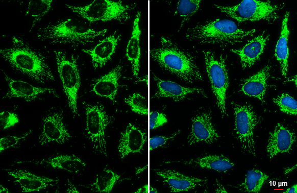
GOT2 antibody detects GOT2 protein at mitochondria by immunofluorescent analysis.Sample: HeLa cells were fixed in ice-cold MeOH for 5 min.Green: GOT2 stained by GOT2 antibody (GTX101930) diluted at 1:500.Blue: Hoechst 33342 staining.Scale bar= 10 μm.
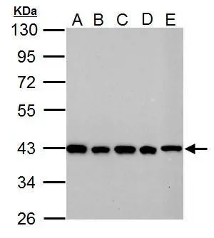
Sample (30 ug of whole cell lysate)
A: NIH-3T3
B: JC
C: BCL-1
D: C2C12
E: Raw264.7
10% SDS PAGE
GTX101930 diluted at 1:10000
-
HostRabbit
-
ClonalityPolyclonal
-
IsotypeIgG
-
ApplicationsWB ICC/IF IHC-P
-
ReactivityHuman, Mouse, Rat