GRP94 antibody


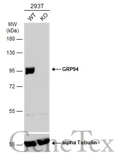
Wild-type (WT) and GRP94 knockout (KO) 293T cell extracts (30 μg) were separated by 7.5% SDS-PAGE, and the membrane was blotted with GRP94 antibody (GTX103232) diluted at 1:2000. The HRP-conjugated anti-rabbit IgG antibody (GTX213110-01) was used to detect the primary antibody.
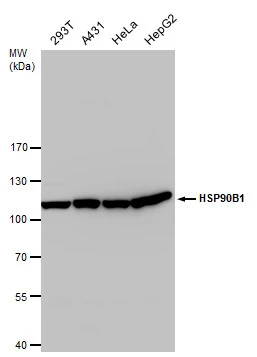
GRP94 antibody detects GRP94 protein by Western blot analysis. Various whole cell extracts (30 μg) were separated by 7.5 % SDS-PAGE, and blotted with GRP94 antibody (GTX103232) diluted by 1:1000
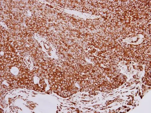
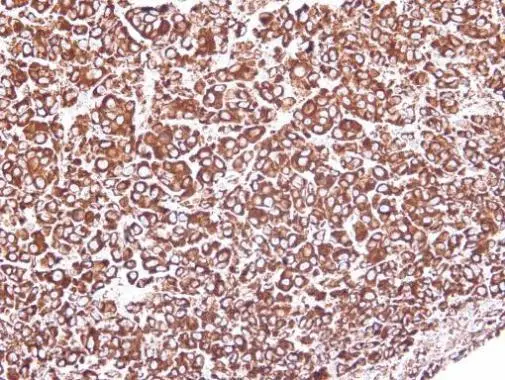
GRP94 antibody detects GRP94 protein at cytoplasm in human lung adenocarcinoma by immunohistochemical analysis.
Sample: Paraffin-embedded human lung adenocarcinoma.
GRP94 antibody (GTX103232) diluted at 1:500.
Antigen Retrieval: Trilogy™ (EDTA based, pH 8.0) buffer, 15min
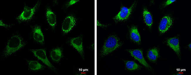
GRP94 antibody detects GRP94 protein at cytoplasm by immunofluorescent analysis.
Sample: HeLa cells were fixed in 4% paraformaldehyde at RT for 15 min.
Green: GRP94 protein stained by GRP94 antibody (GTX103232) diluted at 1:500.
Blue: Hoechst 33342 staining.
Scale bar = 10 μm.
Immunohistochemical analysis of paraffin-embedded HEP3B xenograft, using GRP94(GTX103232) antibody at 1:100 dilution.
Antigen Retrieval: Trilogy™ (EDTA based, pH 8.0) buffer, 15min
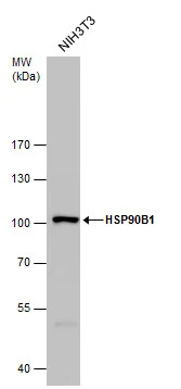
GRP94 antibody detects GRP94 protein by Western blot analysis. Whole cell extracts (30 μg) was separated by 7.5 % SDS-PAGE, and blotted with GRP94 antibody (GTX103232) diluted by 1:1000
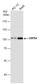
Various whole cell extracts (30 μg) were separated by 7.5% SDS-PAGE, and the membrane was blotted with GRP94 antibody (GTX103232) diluted at 1:1000. The HRP-conjugated anti-rabbit IgG antibody (GTX213110-01) was used to detect the primary antibody.
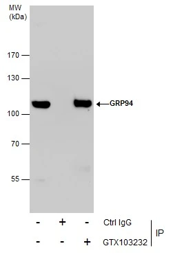
Immunoprecipitation of GRP94 protein from HeLa whole cell extracts using 5 μg of GRP94 antibody (GTX103232).
Western blot analysis was performed using GRP94 antibody (GTX103232).
EasyBlot anti-Rabbit IgG (GTX221666-01) was used as a secondary reagent.
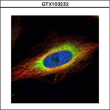
Confocal immunofluorescence analysis (Olympus FV10i) of paraformaldehyde-fixed HeLa, using GRP94(GTX103232) antibody (green) at 1:500 dilution. Alpha-tubulin filaments were labeled with GTX11304 (red) at 1:2500.
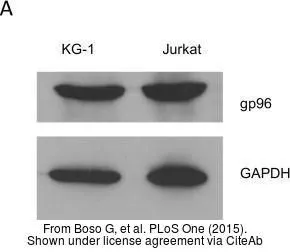
The data was published in the journal PLoS One in 2015. PMID: 25811758
-
HostRabbit
-
ClonalityPolyclonal
-
IsotypeIgG
-
ApplicationsWB ICC/IF IHC-P IP
-
ReactivityHuman, Mouse, Rat