Grp78 antibody


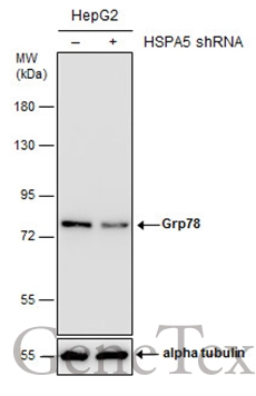
Non-transfected (–) and transfected (+) HepG2 whole cell extracts (30 μg) were separated by 7.5% SDS-PAGE, and the membrane was blotted with Grp78 antibody (GTX113340) diluted at 1:10000. The HRP-conjugated anti-rabbit IgG antibody (GTX213110-01) was used to detect the primary antibody.
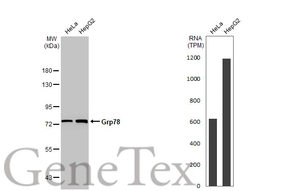
Various whole cell extracts (30 μg) were separated by 7.5% SDS-PAGE, and the membrane was blotted with Grp78 antibody (GTX113340) diluted at 1:10000. The HRP-conjugated anti-rabbit IgG antibody (GTX213110-01) was used to detect the primary antibody. Corresponding RNA expression data for the same cell lines are based on Human Protein Atlas program.
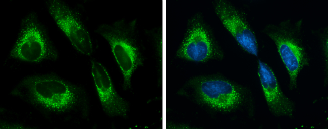
Grp78 antibody detects Grp78 protein at cytoplasm by immunofluorescent analysis.
Sample: HeLa cells were fixed in ice-cold MeOH for 5 min.
Green: Grp78 protein stained by Grp78 antibody (GTX113340) diluted at 1:500.
Blue: Hoechst 33342 staining.
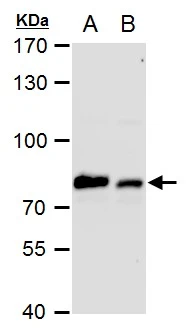
Grp78 antibody detects Grp78 protein by western blot analysis.
A. 30 μg PC-12 whole cell extract
B. 30 μg Rat2 whole cell extract
7.5% SDS-PAGE
Grp78 antibody (GTX113340) dilution: 1:10000
The HRP-conjugated anti-rabbit IgG antibody (GTX213110-01) was used to detect the primary antibody.
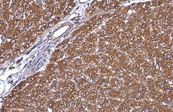
Grp78 antibody detects Grp78 protein at cytoplasm by immunohistochemical analysis.Sample: Paraffin-embedded rat liver.Grp78 stained by Grp78 antibody (GTX113340) diluted at 1:500.Antigen Retrieval: Citrate buffer, pH 6.0, 15 min
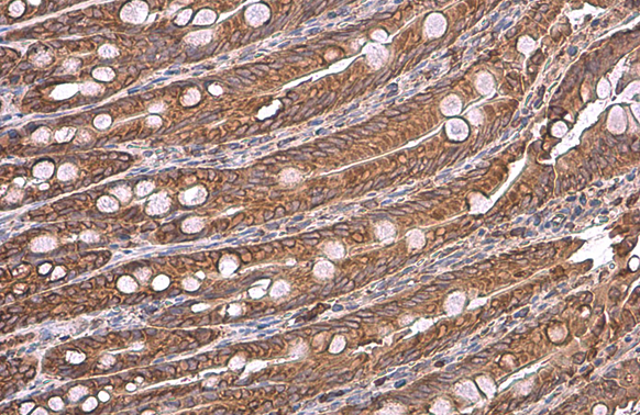
Grp78 antibody detects Grp78 protein at cytoplasm by immunohistochemical analysis.Sample: Paraffin-embedded rat duodenum.Grp78 stained by Grp78 antibody (GTX113340) diluted at 1:500.Antigen Retrieval: Citrate buffer, pH 6.0, 15 min
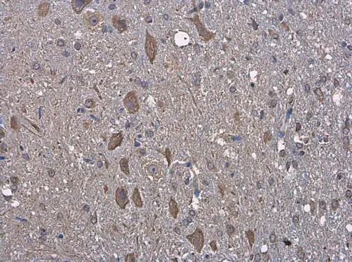
Grp78 antibody detects Grp78 protein at cytoplasm in rat brain by immunohistochemical analysis.
Sample: Paraffin-embedded rat brain.
Grp78 antibody (GTX113340) diluted at 1:250.
Antigen Retrieval: Citrate buffer, pH 6.0, 15 min
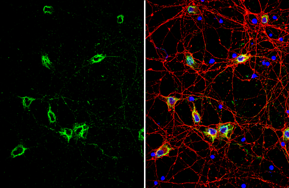
Grp78 antibody detects Grp78 protein by immunofluorescent analysis.Sample: DIV10 rat E18 primary cortical neuron cells were fixed in 4% paraformaldehyde at RT for 15 min.Green: Grp78 stained by Grp78 antibody (GTX113340) diluted at 1:500.Red: Tau, stained by Tau antibody [GT287] (GTX634809) diluted at 1:500.Blue: Fluoroshield with DAPI (GTX30920).
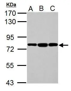
Sample (30 μg of whole cell lysate)
A: NIH-3T3
B: JC
C: BCL-1
7.5% SDS PAGE
GTX113340 diluted at 1:10000
The HRP-conjugated anti-rabbit IgG antibody (GTX213110-01) was used to detect the primary antibody.
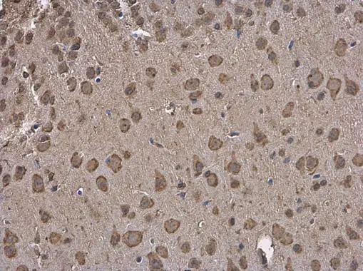
Grp78 antibody detects Grp78 protein at cytoplasm in mouse brain by immunohistochemical analysis.
Sample: Paraffin-embedded mouse brain.
Grp78 antibody (GTX113340) diluted at 1:250.
Antigen Retrieval: Citrate buffer, pH 6.0, 15 min
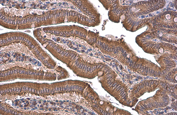
Grp78 antibody detects Grp78 protein at cytoplasm by immunohistochemical analysis.Sample: Paraffin-embedded mouse intestine.Grp78 stained by Grp78 antibody (GTX113340) diluted at 1:500.Antigen Retrieval: Citrate buffer, pH 6.0, 15 min
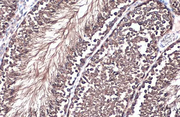
Grp78 antibody detects Grp78 protein at cytoplasm by immunohistochemical analysis.Sample: Paraffin-embedded mouse testis.Grp78 stained by Grp78 antibody (GTX113340) diluted at 1:500.Antigen Retrieval: Citrate buffer, pH 6.0, 15 min
-
HostRabbit
-
ClonalityPolyclonal
-
IsotypeIgG
-
ApplicationsWB ICC/IF IHC-P
-
ReactivityHuman, Mouse, Rat, Chicken