HADH antibody

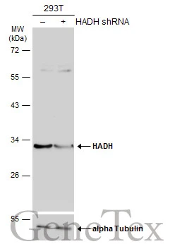
Non-transfected (–) and transfected (+) 293T whole cell extracts (30 μg) were separated by 10% SDS-PAGE, and the membrane was blotted with HADH antibody (GTX105167) diluted at 1:1000. The HRP-conjugated anti-rabbit IgG antibody (GTX213110-01) was used to detect the primary antibody, and the signal was developed with Trident ECL plus-Enhanced.
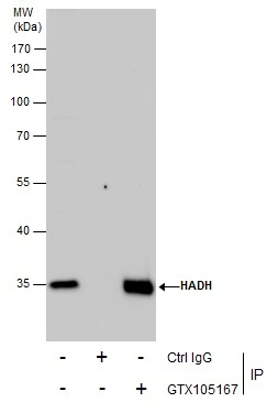
Immunoprecipitation of HADH protein from HepG2 whole cell extracts using 5 μg of HADH antibody (GTX105167).
Western blot analysis was performed using HADH antibody (GTX105167).
EasyBlot anti-Rabbit IgG (GTX221666-01) was used as a secondary reagent.
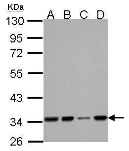
HADH antibody detects HADH protein by Western blot analysis.
A. 30 μg 293T whole cell lysate/extract
B. 30 μg A431 whole cell lysate/extract
C. 30 ug HeLa whole cell lysate/extract
D. 30 ug HepG2 whole cell lysate/extract
10 % SDS-PAGE
HADH antibody (GTX105167) dilution: 1:1000
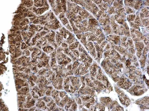
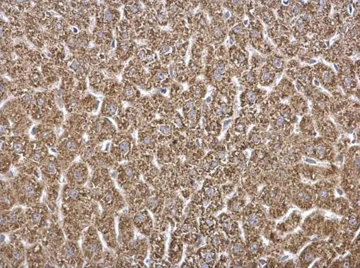
HADH antibody detects HADH protein at mitochondria on mouse liver by immunohistochemical analysis.
Sample: Paraffin-embedded mouse pancreas.
HADH antibody (GTX105167) dilution: 1:500.
Antigen Retrieval: Trilogy™ (EDTA based, pH 8.0) buffer, 15min
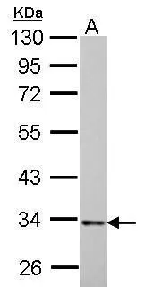
HADH antibody detects HADH protein by Western blot analysis.
A. 30 μg PC-12 whole cell lysate/extract
10 % SDS-PAGE
HADH antibody (GTX105167) dilution: 1:1000
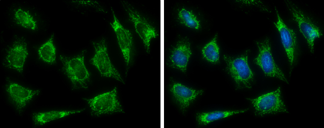
HADH antibody detects HADH protein at mitochondria by immunofluorescent analysis.
Sample: HeLa cells were fixed in ice-cold MeOH for 5 min.
Green: HADH protein stained by HADH antibody (GTX105167) diluted at 1:500.
Blue: Hoechst 33342 staining.
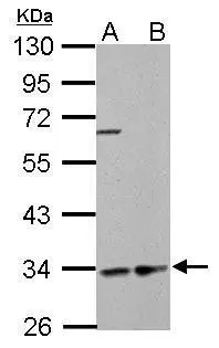
HADH antibody detects HADH protein by Western blot analysis.
A. 30 μg GL261 whole cell lysate/extract
B. 30 ug C8D30 whole cell lysate/extract
10 % SDS-PAGE
HADH antibody (GTX105167) dilution: 1:1000
HADH antibody detects HADH protein at mitochondria on mouse liver by immunohistochemical analysis.
Sample: Paraffin-embedded mouse pancreas.
HADH antibody (GTX105167) dilution: 1:500.
Antigen Retrieval: Trilogy™ (EDTA based, pH 8.0) buffer, 15min
-
HostRabbit
-
ClonalityPolyclonal
-
IsotypeIgG
-
ApplicationsWB ICC/IF IHC-P IP
-
ReactivityHuman, Mouse, Rat