HADH antibody
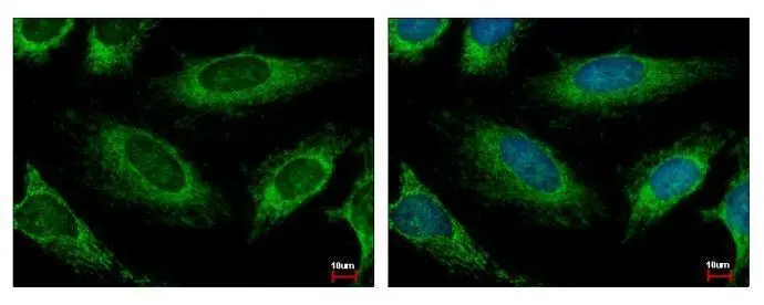
HADH antibody detects HADH protein at Mitochondria by immunofluorescent analysis.
Sample: HeLa cells were fixed in 2% paraformaldehyde/culture medium at 37ºC for 30 min.
Green: HADH protein stained by HADH antibody (GTX118325) diluted at 1:500.
Blue: Hoechst 33343 staining.
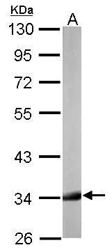
Sample (50 μg of whole cell lysate)
A: mouse liver
10% SDS PAGE
GTX118325 diluted at 1:1000
The HRP-conjugated anti-rabbit IgG antibody (GTX213110-01) was used to detect the primary antibody.
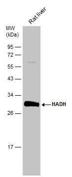
Rat tissue extract (50 μg) was separated by 12% SDS-PAGE, and the membrane was blotted with HADH antibody (GTX118325) diluted at 1:1000. The HRP-conjugated anti-rabbit IgG antibody (GTX213110-01) was used to detect the primary antibody.
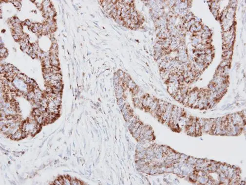
HADH antibody detects HADH protein at mitochondria on human colon carcinoma by immunohistochemical analysis.
Sample: Paraffin-embedded colon carcinoma.
HADH antibody (GTX118325) dilution: 1:250.
Antigen Retrieval: Trilogy™ (EDTA based, pH 8.0) buffer, 15min
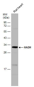
Rat tissue extract (50 μg) was separated by 12% SDS-PAGE, and the membrane was blotted with HADH antibody (GTX118325) diluted at 1:1000. The HRP-conjugated anti-rabbit IgG antibody (GTX213110-01) was used to detect the primary antibody.
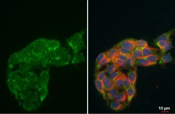
HADH antibody detects HADH protein at mitochondria by immunofluorescent analysis.Sample: HepG2 cells were fixed in 4% paraformaldehyde at RT for 15 min.Green: HADH stained by HADH antibody (GTX118325) diluted at 1:500.Red: alpha Tubulin, a cytoskeleton marker, stained by alpha Tubulin antibody [GT114] (GTX628802) diluted at 1:1000.Blue: Fluoroshield with DAPI (GTX30920).
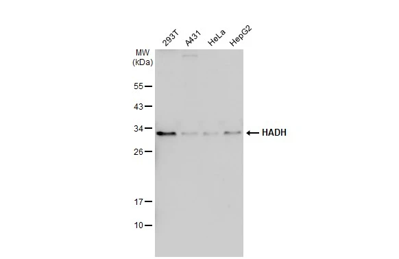
Various whole cell extracts (30 μg) were separated by 12% SDS-PAGE, and the membrane was blotted with HADH antibody (GTX118325) diluted at 1:1000. The HRP-conjugated anti-rabbit IgG antibody (GTX213110-01) was used to detect the primary antibody.
-
HostRabbit
-
ClonalityPolyclonal
-
IsotypeIgG
-
ApplicationsWB ICC/IF IHC-P
-
ReactivityHuman, Mouse, Rat