HAX1 antibody
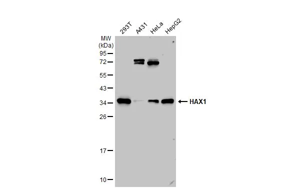
Various whole cell extracts (30 μg) were separated by 12% SDS-PAGE, and the membrane was blotted with HAX1 antibody (GTX101992) diluted at 1:1000. The HRP-conjugated anti-rabbit IgG antibody (GTX213110-01) was used to detect the primary antibody.
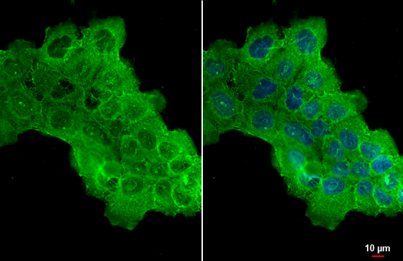
HAX1 antibody detects HAX1 protein at cytoplasm by immunofluorescent analysis.Sample: A431 cells were fixed in ice-cold MeOH for 5 min.Green: HAX1 stained by HAX1 antibody (GTX101992) diluted at 1:500.
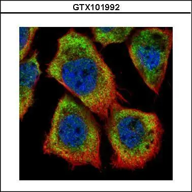
Confocal immunofluorescence analysis (Olympus FV10i) of methanol-fixed A431, using HAX1(GTX101992) antibody (Green) at 1:500 dilution. Alpha-tubulin filaments were labeled with GTX11304 (Red) at 1:2000.
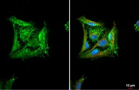
HAX1 antibody detects HAX1 protein at cell membrane by immunofluorescent analysis.Sample: HeLa cells were fixed in ice-cold MeOH for 5 min.Green: HAX1 stained by HAX1 antibody (GTX101992) diluted at 1:500.Red: alpha Tubulin, a cytoskeleton marker, stained by alpha Tubulin antibody [GT114] (GTX628802) diluted at 1:1000.Blue: Fluoroshield with DAPI (GTX30920).
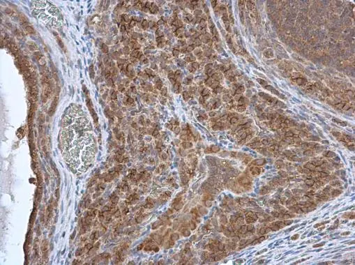
Immunohistochemical analysis of paraffin-embedded NCIN87 Xenograft , using HAX1(GTX101992) antibody at 1:100 dilution.
Antigen Retrieval: Citrate buffer, pH 6.0, 15 min
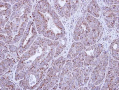
HAX1 antibody detects HAX1 protein at cytoplasm in rat ovary by immunohistochemical analysis.
Sample: Paraffin-embedded rat ovary.
HAX1 antibody (GTX101992) diluted at 1:500.
Antigen Retrieval: Citrate buffer, pH 6.0, 15 min
-
HostRabbit
-
ClonalityPolyclonal
-
IsotypeIgG
-
ApplicationsWB ICC/IF IHC-P
-
ReactivityHuman, Mouse, Rat