PRODUCT
Summary
HIF1 alpha antibody recognizes HIF1 alpha (HIF1a) protein, which is a subunit of the heterodimeric transcription factor hypoxia-inducible factor-1 (HIF-1). HIF-1 is a master regulator of homeostatic adaption to hypoxia (low oxygen supply) in various physiological and pathological conditions including normal development and cancer.
APPLICATION
Application Note
*Optimal dilutions/concentrations should be determined by the researcher.
Application |
Dilution |
1:500-1:3000 |
1:100-1:1000 |
1:100-1:1000 |
Assay dependent |
1:100-1:570 |
Assay dependent |
Not tested in other applications.
Calculated MW
Positive Control
MCF-7 , HeLa(500uM CoCl for 24hr) , NIH-3T3 (500 μM CoCl2 treatment for 24 hr) , HepG2 whole cell lysate/extract (1% O2 treatment for 24hr) , 293T transfected HIF1A , HepG2 ( 200 μM CoCl2 treatment for 24 hr) , HepG2 (500 μM CoCl2 treatment for 24 hr)
Specificity/Sensitivity
Knockdown/Knockout validation was supported by references (PMID:25428606)
Predict Reactivity
Dog, Pig(>80% identity)
PROPERTIES
Form
Liquid
Buffer
1XPBS, 1% BSA, 20% Glycerol (pH7). 0.025% ProClin 300 was added as a preservative.
Storage
Store as concentrated solution. Centrifuge briefly prior to opening vial. For short-term storage (1-2 weeks), store at 4ºC. For long-term storage, aliquot and store at -20ºC or below. Avoid multiple freeze-thaw cycles.
Concentration
0.37 mg/ml (Please refer to the vial label for the specific concentration.)
Antigen Species
Human
Immunogen
Recombinant protein encompassing a sequence within the C-terminus region of human HIF1 alpha. The exact sequence is proprietary.
Purification
Purified by antigen-affinity chromatography.
Conjugation
Unconjugated
RRID
AB_2616089
Note
For laboratory use only. Not for any clinical, therapeutic, or diagnostic use in humans or animals. Not for animal or human consumption.
TARGET
Synonyms
hypoxia inducible factor 1 subunit alpha , HIF-1-alpha , HIF-1A , HIF-1alpha , HIF1 , HIF1-ALPHA , MOP1 , PASD8 , bHLHe78
Cellular Localization
Cytoplasm , Nucleus
Background
Hypoxia-inducible factor-1 (HIF1) is a transcription factor found in mammalian cells cultured under reduced oxygen tension that plays an essential role in cellular and systemic homeostatic responses to hypoxia. HIF1 is a heterodimer composed of an alpha subunit and a beta subunit. The beta subunit has been identified as the aryl hydrocarbon receptor nuclear translocator (ARNT). This gene encodes the alpha subunit of HIF-1. Overexpression of a natural antisense transcript (aHIF) of this gene has been shown to be associated with nonpapillary renal carcinomas. Two alternative transcripts encoding different isoforms have been identified. [provided by RefSeq]
Database
Research Area
DATA IMAGES
|
GTX127309 ICC/IF Image
HIF1 alpha antibody detects HIF1 alpha protein at nucleus in CoCl2-treated samples by confocal immunofluorescent analysis. Samples: Untreated (left panel) and 500μM CoCl2 treated (right panel) HeLa cells for 24 hrs were fixed in 4% paraformaldehyde for 15 min Green: HIF1alpha protein stained by HIF1 alpha antibody (GTX127309) diluted at 1:1000 Red: Alpha-tubulin, a cytoskeleton marker, stained by Alpha-tubulin antibody (GTX11304) diluted at 1:2500 Blue: Hoechst 33342 staining. Scale bar = 10 μm. Images captured by Olympus FV10i Confocal Laser Scanning Microscope
|
|
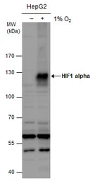
GTX127309 WB Image
HIF1 alpha antibody detects HIF1 alpha protein by western blot analysis. Un-treated (-) and treated (+, 1% O2 treatment for 24hr) HepG2 whole cell extracts (30 μg) were separated by 7.5% SDS-PAGE, and the membrane was blotted with HIF1 alpha antibody (GTX127309) diluted at 1:1000. The HRP-conjugated anti-rabbit IgG antibody (GTX213110-01) was used to detect the primary antibody.
|
|
GTX127309 WB Image
Untreated (–) and treated (+) MCF-7 whole cell extracts (30 μg) were separated by 7.5% SDS-PAGE, and the membrane was blotted with HIF1 alpha antibody (GTX127309) diluted at 1:1000. The HRP-conjugated anti-rabbit IgG antibody (GTX213110-01) was used to detect the primary antibody.
|
|
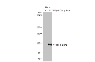
GTX127309 WB Image
Untreated (–) and treated (+) HeLa whole cell extracts (30 μg) were separated by 7.5% SDS-PAGE, and the membrane was blotted with HIF1 alpha antibody (GTX127309) diluted at 1:1000. The HRP-conjugated anti-rabbit IgG antibody (GTX213110-01) was used to detect the primary antibody.
|
|
GTX127309 IHC-P Image
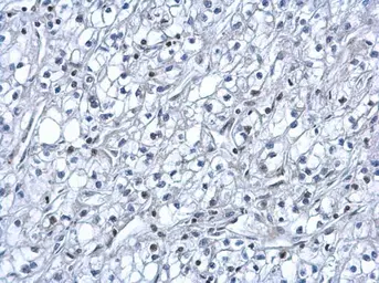
HIF1 alpha antibody detects HIF1 alpha protein at nucleus on human kidney cancer by immunohistochemical analysis. Sample: Paraffin-embedded human kidney cancer. HIF1 alpha antibody (GTX127309) dilution: 1:500.
|
|
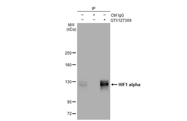
GTX127309 IP Image
Immunoprecipitation of HIF1 alpha protein from HepG2 whole cell extracts treated with 500 μM CoCl2 for 24 hr using 5 μg of HIF1 alpha antibody (GTX127309). Western blot analysis was performed using HIF1 alpha antibody (GTX127309) diluted at 1:500. EasyBlot anti-Rabbit IgG (GTX221666-01) was used as a secondary reagent.
|
|
GTX127309 ChIP assay Image
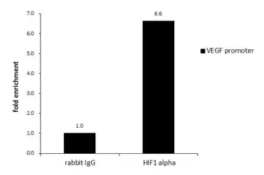
ChIP was performed with HepG2 chromatin extract and 5 μg of either normal rabbit IgG or anti-HIF1 alpha antibody. The precipitated DNA was detected by PCR with primer set targeting to VEGF promoter.
|
|
GTX127309 ICC/IF Image
HIF1 alpha antibody detects HIF1 alpha protein at nucleus by immunofluorescent analysis. Sample: NIH/3T3 cells were fixed in 4% paraformaldehyde at RT for 15 min. Green: HIF1 alpha protein stained by HIF1 alpha antibody (GTX127309) diluted at 1:200. Blue: Hoechst 33342 staining.
|
|
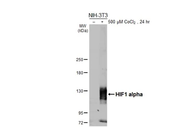
GTX127309 WB Image
Untreated (–) and treated (+) NIH-3T3 whole cell extracts (30 μg) were separated by 7.5% SDS-PAGE, and the membrane was blotted with HIF1 alpha antibody (GTX127309) diluted at 1:500. The HRP-conjugated anti-rabbit IgG antibody (GTX213110-01) was used to detect the primary antibody.
|
|
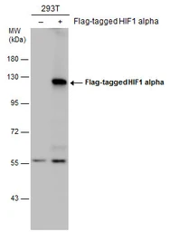
GTX127309 WB Image
Non-transfected (–) and transfected (+) 293T whole cell extracts (30 μg) were separated by 7.5% SDS-PAGE, and the membrane was blotted with HIF1 alpha antibody (GTX127309) diluted at 1:5000. The HRP-conjugated anti-rabbit IgG antibody (GTX213110-01) was used to detect the primary antibody.
|
|
GTX127309 WB Image
Untreated (–) and treated (+) HeLa whole cell extracts (30 μg) were separated by 5% SDS-PAGE, and the membrane was blotted with HIF1 alpha antibody (GTX127309) diluted at 1:1000. The HRP-conjugated anti-rabbit IgG antibody (GTX213110-01) was used to detect the primary antibody.
|
|
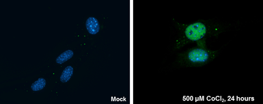
GTX127309 ICC/IF Image
HIF1 alpha antibody detects HIF1 alpha protein at nucleus by immunofluorescent analysis.Sample: Mock and treated HeLa cells were fixed in 4% paraformaldehyde at RT for 15 min.Green: HIF1 alpha stained by HIF1 alpha antibody (GTX127309) diluted at 1:500.Red: phalloidin, a cytoskeleton marker, diluted at 1:200.Scale bar= 10 μm.
|
REFERENCE
REVIEW
HIF1 alpha antibody
Cat No. GTX127309
Rating
|
|
( Average 4.6 based on 42 users reviews)
|
Application
Western Blot(WB)
|
|
( Average 4.8 based on 36 users reviews)
|
Immunocytochemistry/ Immunofluorescence(ICC/IF)
|
|
( Average 4.5 based on 2 users reviews)
|
IHC (Paraffin sections)(IHC-P)
|
|
( Average 3.7 based on 3 users reviews)
|
IHC (Frozen sections)(IHC-Fr)
|
|
( Average 4 based on 1 users reviews)
|
Date :
Anonymous submitted on 23-Dec-2020
Application Tested :
WB
Sample Species :
Hu
Figure Note :
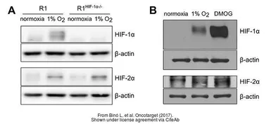
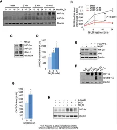
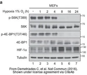
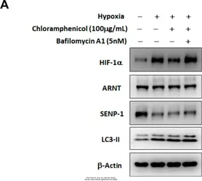
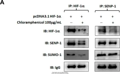
The data was pear reviewed and published in the journal Cells in 2019.
PMID: 31905895
Date :
Anonymous submitted on 23-Dec-2020
Application Tested :
WB
Sample Species :
Hu
Figure Note :
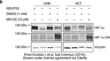
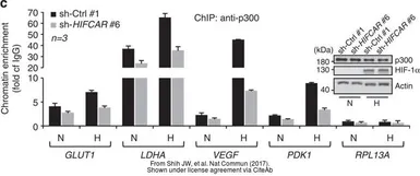
The data was pear reviewed and published in the journal Nat Commun in 2019.
PMID: 30796225
Date :
Anonymous submitted on 23-Dec-2020
Application Tested :
WB
Sample Species :
Hu
Figure Note :
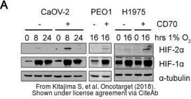
The data was pear reviewed and published in the journal Oncotarget in 2018.
PMID: 29721188
Date :
Anonymous submitted on 23-Dec-2020
Application Tested :
WB
Sample Species :
Hu
Figure Note :
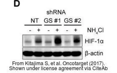
The data was pear reviewed and published in the journal Oncotarget in 2017.
PMID: 29383096
Date :
Anonymous submitted on 23-Dec-2020
Application Tested :
WB
Sample Species :
Ms
Figure Note :
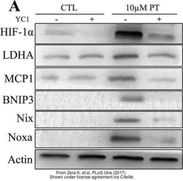
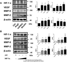
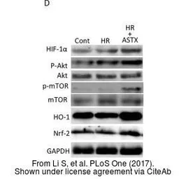
The data was pear reviewed and published in the journal PLoS One in 2017.
PMID: 29045486
Date :
Anonymous submitted on 23-Dec-2020
Application Tested :
WB
Sample Species :
Ms
Figure Note :
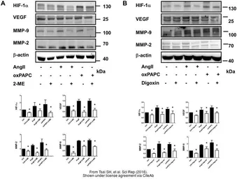
The data was pear reviewed and published in the journal PLoS One in 2017.
PMID: 29045486
Date :
Anonymous submitted on 23-Dec-2020
Application Tested :
ICC/IF
Sample Species :
Hu
Figure Note :
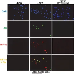
The data was pear reviewed and published in the journal PLoS Pathog in 2017.
PMID: 28617871
Date :
Anonymous submitted on 23-Dec-2020
Application Tested :
WB
Sample Species :
Hu
Figure Note :
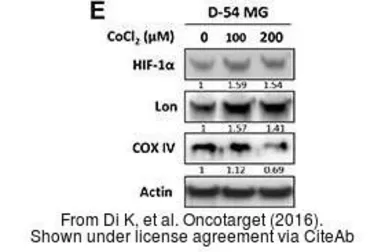
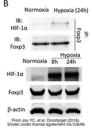
The data was pear reviewed and published in the journal Oncotarget in 2016.
PMID: 27764809
Date :
Anonymous submitted on 23-Dec-2020
Application Tested :
WB
Sample Species :
Hu
Figure Note :
The data was pear reviewed and published in the journal Oncotarget in 2016.
PMID: 27557492
Date :
Anonymous submitted on 23-Dec-2020
Application Tested :
WB
Sample Species :
Hu
Figure Note :
The data was pear reviewed and published in the journal Oncotarget in 2017.
PMID: 29383096
Date :
Anonymous submitted on 23-Dec-2020
Application Tested :
WB
Sample Species :
Ms
Figure Note :
The data was pear reviewed and published in the journal Oncotarget in 2017.
PMID: 29137374
Date :
Anonymous submitted on 23-Dec-2020
Application Tested :
WB
Sample Species :
Ms
Figure Note :
The data was pear reviewed and published in the journal PLoS One in 2017.
PMID: 29045486
Date :
Anonymous submitted on 23-Dec-2020
Application Tested :
WB
Sample Species :
Hu
Figure Note :
The data was pear reviewed and published in the journal Oncotarget in 2016.
PMID: 27557492
Date :
Anonymous submitted on 19-Nov-2020
Application Tested :
WB
Sample Species :
Ms
Figure Note :
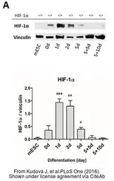
The data was pear reviewed and published in the journal PLoS One in 2016 .
PMID: 27355368
Date :
Anonymous submitted on 19-Nov-2020
Application Tested :
WB
Sample Species :
Hu
Figure Note :
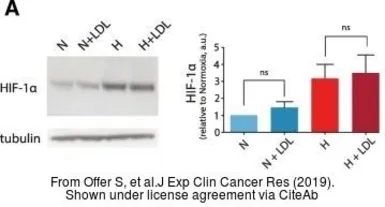
The data was pear reviewed and published in the journal J Exp Clin Cancer Res in 2019 .
PMID: 31174567
Date :
Anonymous submitted on 19-Nov-2020
Application Tested :
WB
Sample Species :
Hu
Figure Note :
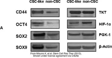
The data was pear reviewed and published in the journal Front Microbiol in 2017.
PMID: 29312159
Date :
Anonymous submitted on 19-Nov-2020
Application Tested :
WB
Sample Species :
Hu
Figure Note :
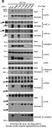
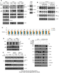
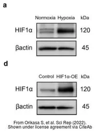
The data was pear reviewed and published in the journal Sci Rep in 2017.
PMID: 28775317
Date :
Anonymous submitted on 19-Nov-2020
Application Tested :
WB
Sample Species :
Hu
Figure Note :
The data was pear reviewed and published in the journal Sci Rep in 2017.
PMID: 28775317
Date :
Anonymous submitted on 19-Nov-2020
Application Tested :
WB
Sample Species :
Hu
Figure Note :
The data was pear reviewed and published in the journal Sci Rep in 2016.
PMID: 27363580
Date :
Anonymous submitted on 19-Nov-2020
Application Tested :
WB
Sample Species :
Hu
Figure Note :
The data was pear reviewed and published in the journal Sci Rep in 2016.
PMID: 27363580
Date :
Anonymous submitted on 19-Nov-2020
Application Tested :
WB
Sample Species :
Hu
Figure Note :
The data was pear reviewed and published in the journal Sci Rep in 2016.
PMID: 27363580
Date :
Anonymous submitted on 19-Nov-2020
Application Tested :
WB
Sample Species :
Hu
Figure Note :
The data was pear reviewed and published in the journal Sci Rep in 2017.
PMID: 28775317
Date :
Anonymous submitted on 19-Nov-2020
Application Tested :
WB
Sample Species :
Hu
Figure Note :
The data was pear reviewed and published in the journal Int J Mol Sci in 2017.
PMID: 28534853
Date :
Anonymous submitted on 19-Nov-2020
Application Tested :
WB
Sample Species :
Ms
Figure Note :
The data was pear reviewed and published in the journal PLoS Negl Trop Dis in 2014.
PMID: 25412261
Date :
Anonymous submitted on 14-Sep-2020
Application Tested :
WB
Sample Species :
Hu
Sample :
LoVo cells
Blocking :
3% skim-milk in TBST, 4°C, 18Hr
Primary Antibody :
1:1000, 25°C, 1Hr
Figure Note :
LoVo cell were cultured using the hypoxic culture kit under hypoxic culture conditions (1% O2 at 37ºC) for 1, 2, 3, or 4 d. Lysates of normoxic and hypoxic cultured cells were separated on 8% SDS-PAGE gels and were immunoblotted using HIF-1 alpha Ab.
Date :
Anonymous submitted on 14-Sep-2020
Application Tested :
WB
Sample Species :
Hu
Sample :
DLD-1 cells
Blocking :
3% skim-milk in TBST, 4°C, 18Hr
Primary Antibody :
1:1000, 25°C, 1Hr
Figure Note :
DLD-1 cell were cultured using the hypoxic culture kit under hypoxic culture conditions (1% O2 at 37ºC) for 1, 2, 3, or 4 d. Lysates of normoxic and hypoxic cultured cells were separated on 8% SDS-PAGE gels and were immunoblotted using HIF-1 alpha Ab.
Date :
Anonymous submitted on 25-Apr-2019
Application Tested :
WB
Sample Species :
Hu
Sample :
RAW264.7 cells
Amount used :
20 μg
Blocking :
3% non-fat dry milk, 25°C, 0.5Hr
Primary Antibody :
1:1000, 25°C, 1Hr
Date :
Anonymous submitted on 14-Feb-2019
Application Tested :
IHC-Fr
Sample Species :
Ms
Sample :
Adult cardiac tissue
Blocking :
10% goat serum in PBS-TX100 0.1%, 25°C, 1Hr
Fixation :
4% PFA
Permeabilization /Antigen retrieval :
0.5% Triton-X-100/Citrate Buffer pH6, 0.15Hr
Primary Antibody :
1:50, 4°C, 16Hr
Date :
Anonymous submitted on 14-Feb-2019
Application Tested :
IHC-P
Sample Species :
Hu
Sample :
Renal clear cell carcinoma
Blocking :
EnVISION FLEX PEROXIDASE-BLOCKING REAGENT (Dako, Ref. K800)
Fixation :
3.7% formalin
Permeabilization /Antigen retrieval :
pH 9 Envision FLEX target retrieval solution, High pH, (Dako Ref. GV800)
Primary Antibody :
1:100, 25°C, 1Hr
Date :
Anonymous submitted on 30-Nov-2017
Application Tested :
WB
Sample Species :
Hu
Sample :
HeLa cells
Amount used :
10 μg
Blocking :
ATTO EzBlock Chemi, 25°C, 0.5Hr
Primary Antibody :
1:5000, 4°C, 16Hr
Date :
Anonymous submitted on 22-Nov-2017
Application Tested :
IHC-P
Sample :
HeLa xenograft
Blocking :
10% goat serum, 25°C, 0.08Hr
Fixation :
Formalin Neutral Buffer
Permeabilization /Antigen retrieval :
0.2% Brij35 in PBS/Dako S2369
Primary Antibody :
1:100, 25°C, 0.6Hr
Date :
Anonymous submitted on 05-Oct-2017
Application Tested :
WB
Sample Species :
Hu
Sample :
OVSAYO cells
Blocking :
3% skim milk, 25°C, 0.5Hr
Primary Antibody :
1:2000, 4°C, 16Hr
Lane Description :
Lane 1: Vehicle, Lane 2: 500 μM Cocl2 treatment for 4 hr
Date :
Anonymous submitted on 05-Oct-2017
Application Tested :
WB
Sample Species :
Hu
Sample :
OVSAYO cells
Blocking :
3% skim milk, 25°C, 0.5Hr
Primary Antibody :
1:2000, 4°C, 16Hr
Lane Description :
Lane 1: Normoxia, Lane 2: 1% O2 treatment for 4 hr
Date :
Anonymous submitted on 22-Sep-2016
Application Tested :
WB
Sample Species :
Hu
Sample :
dental pulp stem cells
Amount used :
1x10^4 cells
Blocking :
PVDF blocking reagent, 25°C, 0.25Hr
Primary Antibody :
1:1000, 4°C, 16Hr
Date :
Anonymous submitted on 02-Mar-2016
Application Tested :
WB
Sample Species :
Hu
Sample :
THP-1 cells
Amount used :
20μg
Blocking :
5% milk in TBST, 25°C, 1Hr
Primary Antibody :
1:2000, 4°C, 16Hr
Lane Description :
Lane 1:THP-1 cells (250ug/ml TPA treatment for 4h), Lane 2:THP-1 cells (250ug/ml TPA and 1 ug/ml LPS treatment for 4h)
Date :
Anonymous submitted on 29-Jan-2016
Application Tested :
ICC/IF
Sample Species :
Hu
Sample :
A549 cells
Fixation :
4% PFA, 2Hr
Permeabilization /Antigen retrieval :
Citrate buffer, 95°C, 20minHr
Primary Antibody :
1:1000, 25°C, 1Hr
Figure Note :
A: A549 cells, B: A549 cells (1mM DMOG treatment for 3.5 h)
Date :
Anonymous submitted on 29-Jan-2016
Application Tested :
IHC-P
Sample Species :
Hu
Sample :
brain tissue
Fixation :
4% PFA, 2Hr
Permeabilization /Antigen retrieval :
Citrate buffer, 95°C, 20minHr
Primary Antibody :
1:1000, 25°C, 1Hr
Figure Note :
A:Human glioblastoma tissue, B:Human healthy brain tissue
Date :
Anonymous submitted on 30-Dec-2015
Application Tested :
WB
Sample Species :
Hu
Sample :
meniscal fibrochondrocytes
Amount used :
15μg
Blocking :
BlockingPRO Blocking buffer, 25°C, 1Hr
Primary Antibody :
1:1000, 4°C, 16Hr
Lane Description :
Lane 1: meniscal fibrochondrocytes (normoxia), Lane 2: meniscal fibrochondrocytes (1% O2 treatment for 6hr)
Date :
Anonymous submitted on 08-Dec-2014
Application Tested :
WB
Sample Species :
Hu
Sample :
endometriotic stromal cells
Amount used :
20μg
Blocking :
5% non-fat milk, 25°C, 1Hr
Primary Antibody :
1:5000, 25°C, 1Hr
Lane Description :
Lane A:DMSO treatment (24hr) ; Lane B:0.1uM DFO treatment (24hr) ; Lane C:1uM DFO treatment (24hr)
Date :
Anonymous submitted on 14-Aug-2014
Application Tested :
WB
Sample Species :
Hu
Sample :
HeLa cells
Amount used :
20μg
Blocking :
25°C, 1Hr
Primary Antibody :
1:1000, 4°C, 12Hr
Lane Description :
Lane A: HeLa cell lysate(normoxia), Lane B: HeLa cell lysate (500μM CoCl2 treatment)
Date :
Anonymous submitted on 27-Aug-2013
Application Tested :
WB
Sample Species :
Hu
Sample :
A431 cells
Amount used :
20μg
Blocking :
5% milk in TBST, 37°C, 1Hr
Primary Antibody :
1:1000, 37°C, 1Hr
Lane Description :
A: A431 whole cell lysate under normoxia, B: A431 whole cell lysate under hypoxia
Date :
Anonymous submitted on 16-Oct-2012
Application Tested :
WB
Sample Species :
Hu
Sample :
A549 cells
Amount used :
40μg
Blocking :
3% milk , 25°C, 1Hr
Primary Antibody :
1:10000, 4°C, 16Hr