HMGB1 + HMGB2 antibody

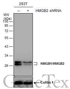
Non-transfected (–) and transfected (+) 293T whole cell extracts (30 μg) were separated by 12% SDS-PAGE, and the membrane was blotted with HMGB1/HMGB2 antibody (GTX127972) diluted at 1:20000.
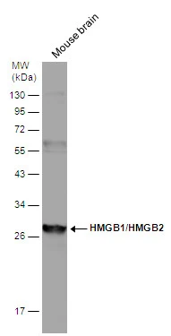
Mouse tissue extract (50 μg) was separated by 12% SDS-PAGE, and the membrane was blotted with HMGB1/HMGB2 antibody (GTX127972) diluted at 1:1000.
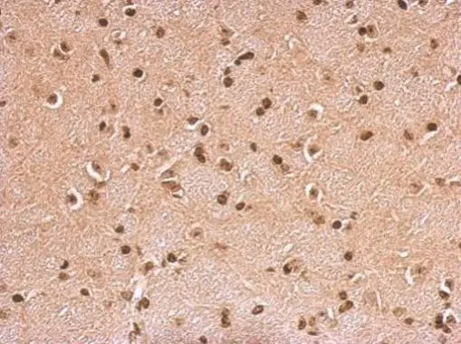
HMGB1/HMGB2 antibody detects HMGB2 protein at cytosol on mouse brain by immunohistochemical analysis.
Sample: Paraffin-embedded mouse brain.
HMGB1/HMGB2 antibody (GTX127972) dilution: 1:500.
Antigen Retrieval: Trilogy™ (EDTA based, pH 8.0) buffer, 15min
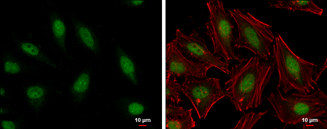
HMGB1/HMGB2 antibody detects HMGB1/HMGB2 protein at nucleus by immunofluorescent analysis.
Sample: HeLa cells were fixed in 4% paraformaldehyde at RT for 15 min.
Green: HMGB1/HMGB2 protein stained by HMGB1/HMGB2 antibody (GTX127972) diluted at 1:2000.
Red: phalloidin, a cytoskeleton marker, diluted at 1:200.
Blue: Hoechst 33342 staining.
Scale bar = 10 μm.
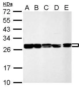
HMGB1/HMGB2 antibody detects HMGB2 protein by Western blot analysis.
A. 30 μg 293T whole cell lysate/extract
B. 30 μg A431 whole cell lysate/extract
C. 30 μg HeLa whole cell lysate/extract
D. 30 μg HepG2 whole cell lysate/extract
E. 30 μg A375 whole cell lysate/extract
12 % SDS-PAGE
HMGB1/HMGB2 antibody (GTX127972) dilution: 1:10000
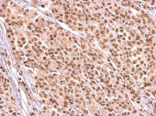
HMGB1/HMGB2 antibody detects HMGB2 protein at nucleus on human duodenal adenocarcinoma tissue by immunohistochemical analysis.
Sample: Paraffin-embedded human duodenal adenocarcinoma tissue.
HMGB1/HMGB2 antibody (GTX127972) dilution: 1:500.
Antigen Retrieval: Trilogy™ (EDTA based, pH 8.0) buffer, 15min
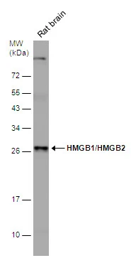
Rat tissue extract (50 μg) was separated by 12% SDS-PAGE, and the membrane was blotted with HMGB1/HMGB2 antibody (GTX127972) diluted at 1:2000.
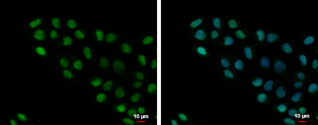
HMGB1/HMGB2 antibody detects HMGB1/HMGB2 protein at nucleus by immunofluorescent analysis.
Sample: A375 cells were fixed in 4% paraformaldehyde at RT for 15 min.
Green: HMGB1/HMGB2 protein stained by HMGB1/HMGB2 antibody (GTX127972) diluted at 1:500.
Blue: Hoechst 33342 staining.
Scale bar = 10 μm.
-
HostRabbit
-
ClonalityPolyclonal
-
IsotypeIgG
-
ApplicationsWB ICC/IF IHC-P
-
ReactivityHuman, Mouse, Rat