HMGB1 antibody


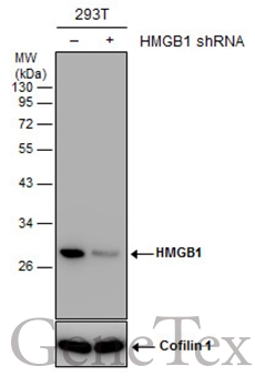
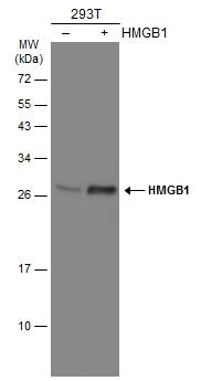
Non-transfected (–) and transfected (+) 293T whole cell extracts (30 μg) were separated by 12% SDS-PAGE, and the membrane was blotted with HMGB1 antibody (GTX112959) diluted at 1:2000. The HRP-conjugated anti-rabbit IgG antibody (GTX213110-01) was used to detect the primary antibody.
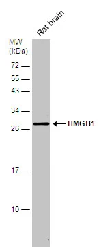
Rat tissue extract (50 μg) was separated by 12% SDS-PAGE, and the membrane was blotted with HMGB1 antibody (GTX112959) diluted at 1:500. The HRP-conjugated anti-rabbit IgG antibody (GTX213110-01) was used to detect the primary antibody.
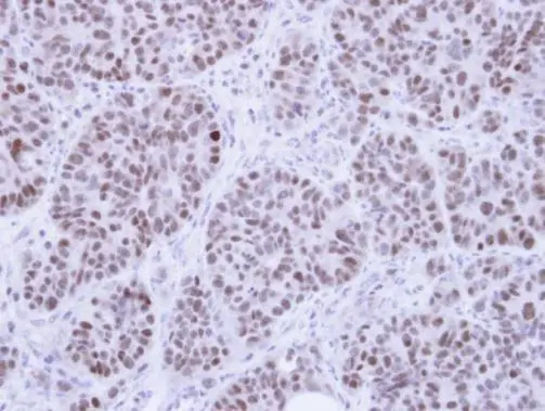
Immunohistochemical analysis of paraffin-embedded SW480 Xenograft, using HMG-1(GTX112959) antibody at 1:100 dilution.
Antigen Retrieval: Trilogy™ (EDTA based, pH 8.0) buffer, 15min
Sample (30 μg of whole cell lysate)
A: HepG2 (GTX27900)
12% SDS PAGE
GTX112959 diluted at 1:1000
The HRP-conjugated anti-rabbit IgG antibody (GTX213110-01) was used to detect the primary antibody.
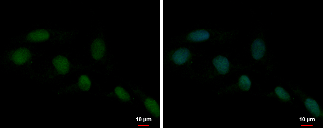
HMGB1 antibody detects HMGB1 protein at nucleus by immunofluorescent analysis.
Sample: NT2D1 cells were fixed in 4% paraformaldehyde at RT for 15 min.
Green: HMGB1 protein stained by HMGB1 antibody (GTX112959) diluted at 1:1000.
Blue: Hoechst 33342 staining.
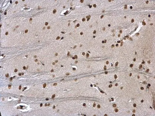
HMGB1 antibody detects HMGB1 protein at nucleus on rat fore brain by immunohistochemical analysis.
Sample: Paraffin-embedded rat fore brain.
HMGB1 antibody (GTX112959) dilution: 1:500.
Antigen Retrieval: Trilogy™ (EDTA based, pH 8.0) buffer, 15min
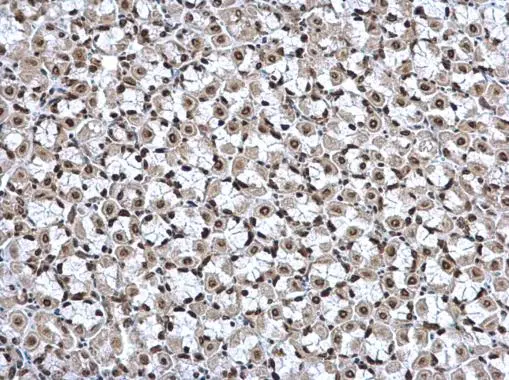
HMGB1 antibody detects HMGB1 protein at nucleus on mouse stomach by immunohistochemical analysis.
Sample: Paraffin-embedded mouse stomach.
HMGB1 antibody (GTX112959) dilution: 1:500.
Antigen Retrieval: Trilogy™ (EDTA based, pH 8.0) buffer, 15min
Various whole cell extracts (30 μg) were separated by 12% SDS-PAGE, and the membrane was blotted with HMGB1 antibody (GTX112959) diluted at 1:500. The HRP-conjugated anti-rabbit IgG antibody (GTX213110-01) was used to detect the primary antibody.
Non-transfected (–) and transfected (+) 293T whole cell extracts (30 μg) were separated by 12% SDS-PAGE, and the membrane was blotted with HMGB1 antibody (GTX112959) diluted at 1:3000. The HRP-conjugated anti-rabbit IgG antibody (GTX213110-01) was used to detect the primary antibody.
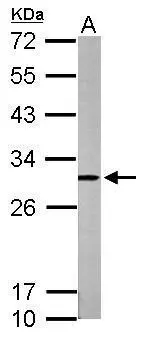
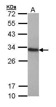
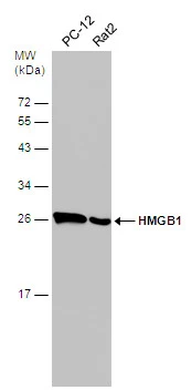
Sample (50 μg of whole cell lysate)
A: mouse brain
12% SDS PAGE
GTX112959 diluted at 1:1000
The HRP-conjugated anti-rabbit IgG antibody (GTX213110-01) was used to detect the primary antibody.
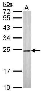
Sample (30 μg of whole cell lysate)
A: whole zebrafish
12% SDS PAGE
GTX112959 diluted at 1:1000
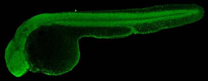
HMGB1 antibody detects Hmgb1 protein on zebrafish by whole mount immunohistochemical analysis.
Sample: 2 days-post-fertilization zebrafish embryo.
HMGB1 antibody (GTX112959) dilution: 1:100.
-
HostRabbit
-
ClonalityPolyclonal
-
IsotypeIgG
-
ApplicationsWB ICC/IF IHC-P IHC-Wm
-
ReactivityHuman, Mouse, Rat, Zebrafish