HPRT antibody


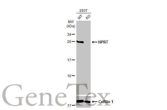
Wild-type (WT) and HPRT knockout (KO) 293T cell extracts (10 μg) were separated by 12% SDS-PAGE, and the membrane was blotted with HPRT antibody (GTX113466) diluted at 1:1000. The HRP-conjugated anti-rabbit IgG antibody (GTX213110-01) was used to detect the primary antibody.
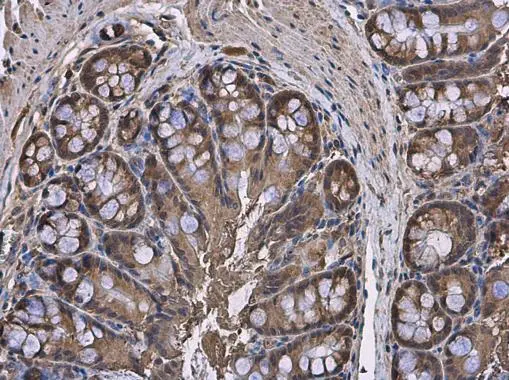
HPRT antibody detects HPRT protein at cytoplasm in rat colon by immunohistochemical analysis.
Sample: Paraffin-embedded rat colon.
HPRT antibody (GTX113466) diluted at 1:500.
Antigen Retrieval: Citrate buffer, pH 6.0, 15 min
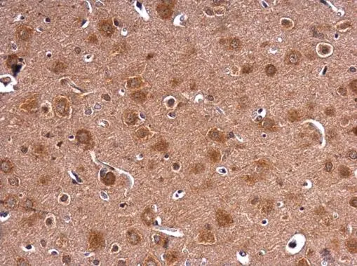
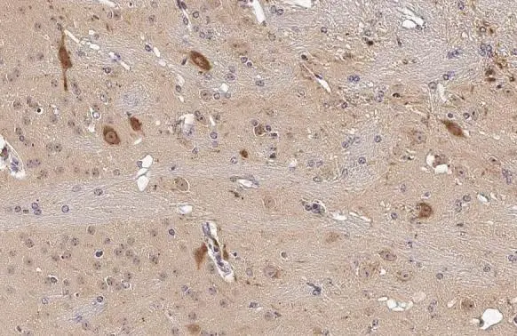
HPRT antibody detects HPRT protein at cytoplasm in rat brain by immunohistochemical analysis.
Sample: Paraffin-embedded rat brain.
HPRT antibody (GTX113466) diluted at 1:500.
Antigen Retrieval: Citrate buffer, pH 6.0, 15 min
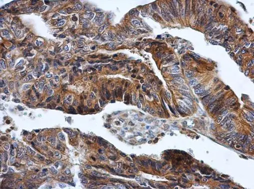
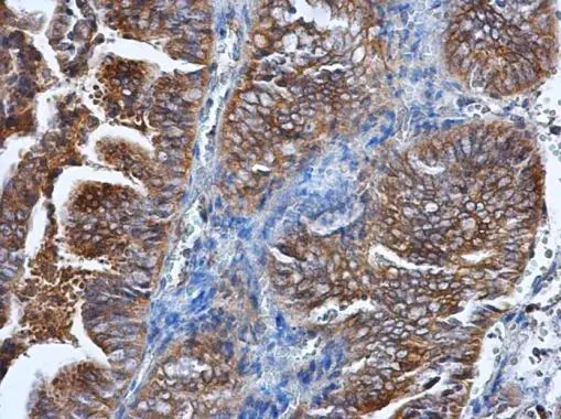
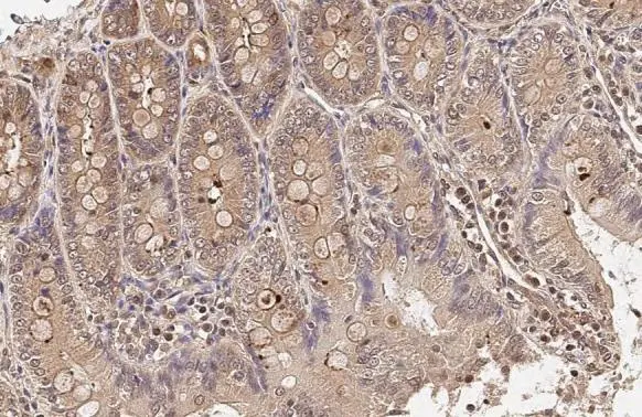
HPRT antibody detects HPRT protein at cytoplasm in human colon cancer by immunohistochemical analysis.
Sample: Paraffin-embedded human colon cancer.
HPRT antibody (GTX113466) diluted at 1:500.
Antigen Retrieval: Citrate buffer, pH 6.0, 15 min
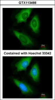
Immunofluorescence analysis of paraformaldehyde-fixed HeLa, using HPRT(GTX113466) antibody at 1:200 dilution.
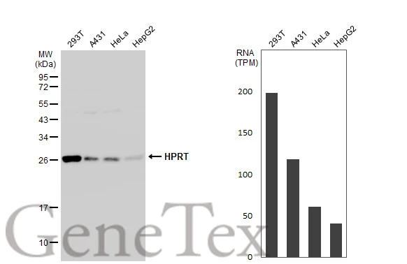
Various whole cell extracts (30 μg) were separated by 12% SDS-PAGE, and the membrane was blotted with HPRT antibody (GTX113466) diluted at 1:500. The HRP-conjugated anti-rabbit IgG antibody (GTX213110-01) was used to detect the primary antibody. Corresponding RNA expression data for the same cell lines are based on Human Protein Atlas program.
HPRT antibody detects HPRT protein at cytoplasm in human endometrial cancer by immunohistochemical analysis.
Sample: Paraffin-embedded human endometrial cancer.
HPRT antibody (GTX113466) diluted at 1:500.
Antigen Retrieval: Citrate buffer, pH 6.0, 15 min
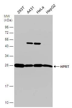
Various whole cell extracts (30 μg) were separated by 12% SDS-PAGE, and the membrane was blotted with HPRT antibody (GTX113466) diluted at 1:1000. The HRP-conjugated anti-rabbit IgG antibody (GTX213110-01) was used to detect the primary antibody.
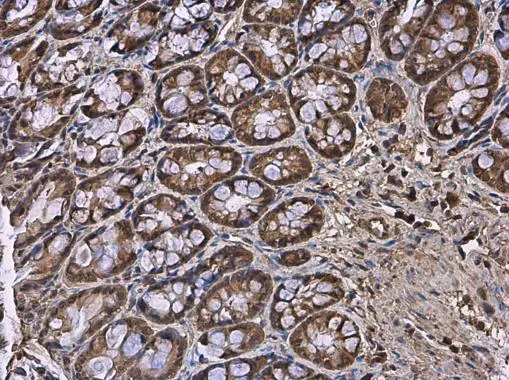
HPRT antibody detects HPRT protein at cytoplasm in mouse colon by immunohistochemical analysis.
Sample: Paraffin-embedded mouse colon.
HPRT antibody (GTX113466) diluted at 1:500.
Antigen Retrieval: Citrate buffer, pH 6.0, 15 min
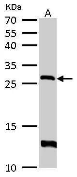
HPRT antibody detects HPRT1 protein by western blot analysis.
A. 50 μg mouse liver lysate/extract
12% SDS-PAGE
HPRT antibody (GTX113466) dilution: 1:500
The HRP-conjugated anti-rabbit IgG antibody (GTX213110-01) was used to detect the primary antibody.
HPRT antibody detects HPRT protein at cytoplasm by immunohistochemical analysis.Sample: Paraffin-embedded mouse brain.HPRT stained by HPRT antibody (GTX113466) diluted at 1:500.Antigen Retrieval: Citrate buffer, pH 6.0, 15 min
HPRT antibody detects HPRT protein at cytoplasm by immunohistochemical analysis.Sample: Paraffin-embedded rat duodenum.HPRT stained by HPRT antibody (GTX113466) diluted at 1:500.Antigen Retrieval: Citrate buffer, pH 6.0, 15 min
-
HostRabbit
-
ClonalityPolyclonal
-
IsotypeIgG
-
ApplicationsWB ICC/IF IHC-P
-
ReactivityHuman, Mouse, Rat