HSP27 antibody

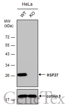
Wild-type (WT) and HSP27 knockout (KO) HeLa cell extracts (30 μg) were separated by 12% SDS-PAGE, and the membrane was blotted with HSP27 antibody (GTX112964) diluted at 1:20000. The HRP-conjugated anti-rabbit IgG antibody (GTX213110-01) was used to detect the primary antibody.
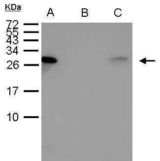
HSP27 antibody immunoprecipitates HSP27 protein in IP experiments. IP Sample: 1000 μg HeLa whole cell lysate/extract A. 40 μg HeLa whole cell lysate/extract B. Control with 2.5 μg of preimmune rabbit IgG C. Immunoprecipitation of HSP27 protein by 2.5 μg of HSP27 (GTX112964) 15% SDS-PAGE The immunoprecipitated HSP27 protein was detected by HSP27 antibody (GTX112964) diluted at 1:1000. EasyBlot anti-rabbit IgG (GTX221666-01) was used as a secondary reagent.
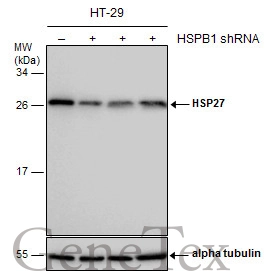
Non-transfected (–) and transfected (+) HT-29 whole cell extracts (30 μg) were separated by 12% SDS-PAGE, and the membrane was blotted with HSP27 antibody (GTX112964) diluted at 1:5000.
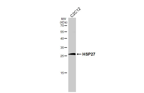
Whole cell extract (30 μg) was separated by 12% SDS-PAGE, and the membrane was blotted with HSP27 antibody (GTX112964) diluted at 1:1000. The HRP-conjugated anti-rabbit IgG antibody (GTX213110-01) was used to detect the primary antibody.

Sample (30 ug of whole cell lysate)
A: 293T
B: A431
C: HeLa
D: HepG2
12% SDS PAGE
GTX112964 diluted at 1:1000
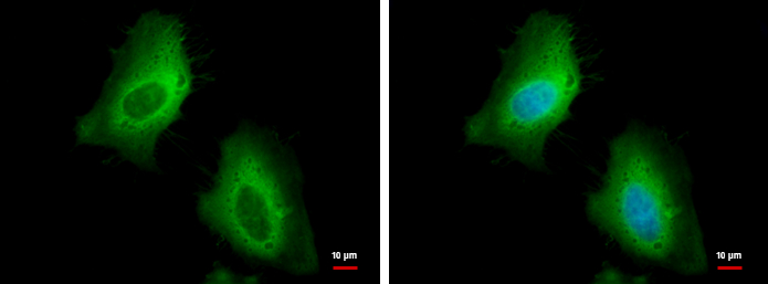
HSP27 antibody detects HSP27 protein at cytoplasm by immunofluorescent analysis.
Sample: HeLa cells were fixed in 4% paraformaldehyde at RT for 15 min.
Green: HSP27 protein stained by HSP27 antibody (GTX112964) diluted at 1:500.
Blue: Hoechst 33342 staining.
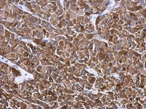
Immunohistochemical analysis of paraffin-embedded U87 xenograft, using HSP27(GTX112964) antibody at 1:500 dilution.
Antigen Retrieval: Trilogy™ (EDTA based, pH 8.0) buffer, 15min
-
HostRabbit
-
ClonalityPolyclonal
-
IsotypeIgG
-
ApplicationsWB ICC/IF IHC-P IP
-
ReactivityHuman, Mouse