APPLICATION
Application Note
IHC: Use at an assay dependent dilution. WB: 1:2000. Detects a band of approximately 32 kDa (predicted molecular weight: 34.6 kDa). Optimal dilutions/concentrations should be determined by the end user.
Calculated MW
Positive Control
Recombinant Human and Rat HO-1 (Hsp32) Protein
Specificity/Sensitivity
May cross-react with HO-2 in western blot. It also recognizes a 50kD band found only in transformed cell lines but not in normal tissues.
PROPERTIES
Form
Liquid
Buffer
Phosphate-buffered saline, pH 7.2, containing 50% glycerol and 0.09% sodium azide
Storage
Store as concentrated solution. Centrifuge briefly prior to opening vial. For short-term storage (1-2 weeks), store at 4ºC. For long-term storage, aliquot and store at -20ºC or below. Avoid multiple freeze-thaw cycles.
Concentration
1mg/ml(Please refer to the vial label for the specific concentration.)
Antigen Species
Rat
Immunogen
Recombinant rat HO-1 (Hsp32) lacking the membrane spanning region
Purification
Protein A affinity purified
From polyclonal serum
Conjugation
Unconjugated
Note
For laboratory use only. Not for any clinical, therapeutic, or diagnostic use in humans or animals. Not for animal or human consumption.
TARGET
Synonyms
Heme Oxygenase 1, Ho1, Heox, 141250, hsp32, 3162, Heme Oxygenase1, P09601, Hmox1, Hmox, HO-1, bK286B10, HEOXG, HO 1
Background
Heme oxygenase (HO) is a microsomal enzyme that catalyzes the oxidation of heme to the antioxidant molecules, biliverdin and carbon monoxide. HO consists of two homologous isozymes, an inducible HO1 and a constitutively expressed HO2. HO1 is induced by a wide variety of stimuli including conditions of oxidative stress, inflammatory agents, transforming growth factor beta and heat shock. The increase in expression of HO1 is thought to be a cellular defense mechanism against oxidative stress since elevated HO could eventually generate more bilirubin, an anti-oxidant.
Database
Research Area
DATA IMAGES
|
GTX30748 IHC-P Image
Immunohistochemistry analysis of human spleen tissue stained with HO-1, pAb at 10µg/ml.
|
|
GTX30748 IHC Image
Immunohistochemistry analysis of human spleen tissue stained with HO-1, pAb at 10µg/ml.
|
|
GTX30748 WB Image
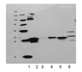
Western blot analysis of HO-1 pAb:
Lane 1: HO-1 Recombinant Protein
Lane 2: Human Liver Microsomes
Lane 3: Rat Liver Microsomes
Lane 4: Mouse Liver Microsomes
Lane 5: Canine Liver Microsomes
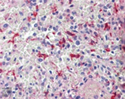
|
|
GTX30748 IHC Image
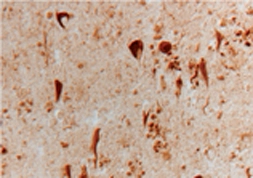
Immunohistochemistry analysis of Alzheimer's diseased section stained with HO-1 (Hsp32) pAb.
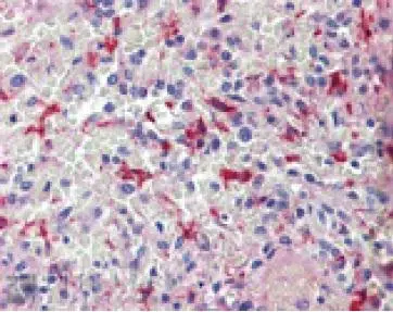
|