Her2 / ErbB2 antibody


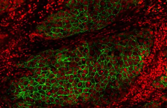
Her2 / ErbB2 antibody detects Her2 / ErbB2 protein at cell membrane by immunohistochemical analysis.Sample: Paraffin-embedded human breast carcinoma.Green: Her2 / ErbB2 stained by Her2 / ErbB2 antibody (GTX100509) diluted at 1:50.
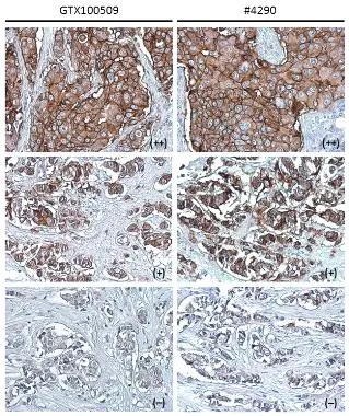
*The competitor is not affiliated with GeneTex and does not endorse this product.
Her2 / ErbB2 antibody [C2C3], C-term detects Her2 / ErbB2 protein at cell membrane in human breast carcinoma by immunohistochemical analysis.
Sample: Strong positive (++), low positive (+) and negative tissue slides cores assessed using Quantitative Digital Pathology.
Her2 / ErbB2 antibody [C2C3], C-term (GTX100509) diluted at 1:500, and competitor's antibody (CST#4290) diluted at 1:100.
Antigen Retrieval: Trilogy™ (EDTA based, pH 8.0) buffer, 15min
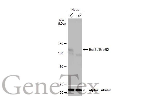
Wild-type (WT) and Her2 / ErbB2 knockout (KO) HeLa cell extracts (30 μg) were separated by 5% SDS-PAGE, and the membrane was blotted with Her2 / ErbB2 antibody (GTX100509) diluted at 1:5000. The HRP-conjugated anti-rabbit IgG antibody (GTX213110-01) was used to detect the primary antibody, and the signal was developed with Trident femto Western HRP Substrate.
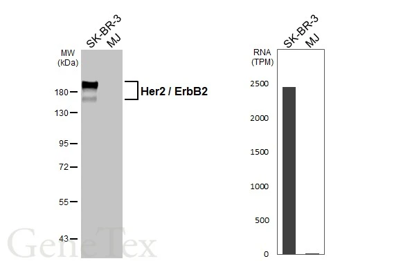
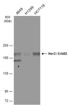
Various whole cell extracts (30 μg) were separated by 7.5% SDS-PAGE, and the membrane was blotted with Her2 / ErbB2 antibody (GTX100509) diluted at 1:1000. The HRP-conjugated anti-rabbit IgG antibody (GTX213110-01) was used to detect the primary antibody. Corresponding RNA expression data for the same cell lines are based on Human Protein Atlas program.
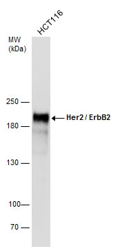
Whole cell extract (30 μg) was separated by 5% SDS-PAGE, and the membrane was blotted with Her2 / ErbB2 antibody [C2C3], C-term (GTX100509) diluted at 1:10000.
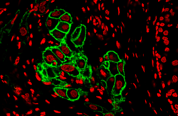
Her2 / ErbB2 antibody detects Her2 / ErbB2 protein at cell membrane by immunohistochemical analysis.
Sample: Human Breast Cancer.
Green: Her2 / ErbB2 stained by Dylight488-conjugated Her2 / ErbB2 antibody (GTX100509) diluted at 1:500.
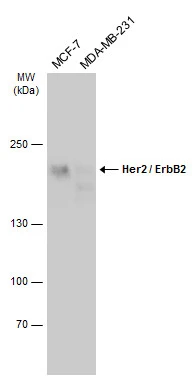
Various whole cell extracts (30 μg) were separated by 5% SDS-PAGE, and the membrane was blotted with Her2 / ErbB2 antibody [C2C3], C-term (GTX100509) diluted at 1:2000. The HRP-conjugated anti-rabbit IgG antibody (GTX213110-01) was used to detect the primary antibody.
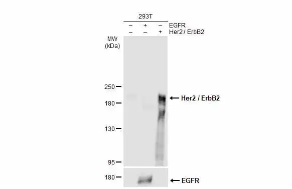
Non-transfected (–) and transfected (+) 293T whole cell extracts were separated by 5% SDS-PAGE, and the membrane was blotted with Her2 / ErbB2 antibody (GTX100509) diluted at 1:7000. The HRP-conjugated anti-rabbit IgG antibody (GTX213110-01) was used to detect the primary antibody.
Various whole cell extracts (30 μg) were separated by 5% SDS-PAGE, and the membrane was blotted with Her2 / ErbB2 antibody [C2C3], C-term (GTX100509) diluted at 1:2000. The HRP-conjugated anti-rabbit IgG antibody (GTX213110-01) was used to detect the primary antibody.
-
HostRabbit
-
ClonalityPolyclonal
-
IsotypeIgG
-
ApplicationsWB IHC-P
-
ReactivityHuman