Hexokinase 1 antibody

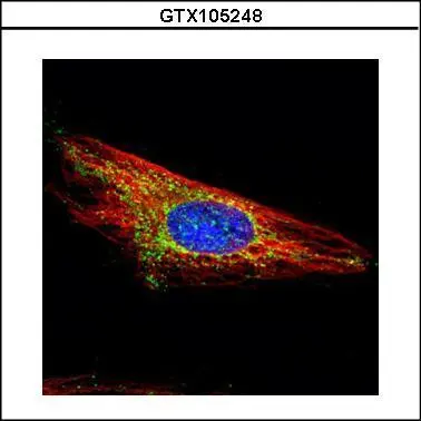
Confocal immunofluorescence analysis (Olympus FV10i) of methanol-fixed HeLa, using HXK I(GTX105248) antibody (Green) at 1:500 dilution. Alpha-tubulin filaments were labeled with GTX11304 (Red) at 1:2000.
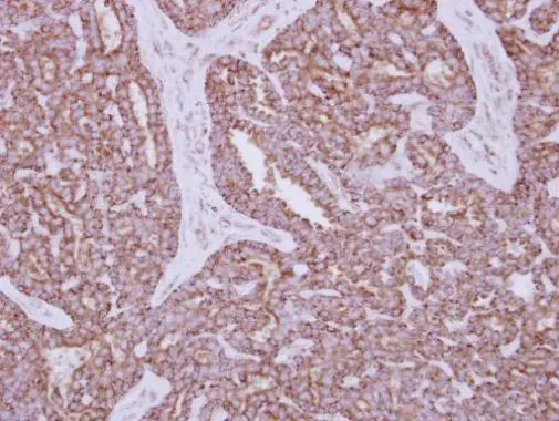
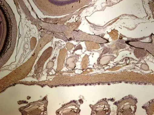
Immunohistochemical analysis of paraffin-embedded human breast cancer, using HXK I(GTX105248) antibody at 1:250 dilution.
Antigen Retrieval: Trilogy™ (EDTA based, pH 8.0) buffer, 15min
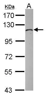
Sample (30 μg of whole cell lysate)
C: zebrafish muscle
7.5% SDS PAGE
GTX105248 diluted at 1:1000
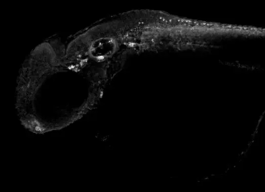
Hexokinase 1 antibody detects Hexokinase 1 protein on zebrafish by whole mount immunohistochemical analysis.
Sample: 2 days-post-fertilization zebrafish embryo.
Hexokinase 1 antibody (GTX105248) dilution: 1:100.
Immunohistochemical analysis of paraffin-embedded zebrafish tissue, using Hexokinase 1 antibody (GTX105248) at 1:300 dilution.
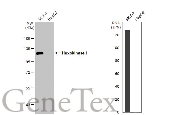
Various whole cell extracts (30 μg) were separated by 7.5% SDS-PAGE, and the membrane was blotted with Hexokinase 1 antibody (GTX105248) diluted at 1:3000. The HRP-conjugated anti-rabbit IgG antibody (GTX213110-01) was used to detect the primary antibody. Corresponding RNA expression data for the same cell lines are based on Human Protein Atlas program.
-
HostRabbit
-
ClonalityPolyclonal
-
IsotypeIgG
-
ApplicationsWB ICC/IF IHC-P IHC-Wm
-
ReactivityHuman, Zebrafish