Histone H2A.X antibody

Histone H2A.X antibody detects Histone H2A.X protein at nucleus on mouse prostate by immunohistochemical analysis.
Sample: Paraffin-embedded mouse prostate.
Histone H2A.X antibody (GTX108272) dilution: 1:500.
Antigen Retrieval: Trilogy™ (EDTA based, pH 8.0) buffer, 15min
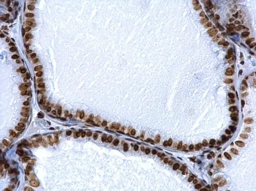
Histone H2A.X antibody detects Histone H2A.X protein at nucleus on rat hind brain by immunohistochemical analysis.
Sample: Paraffin-embedded rat hind brain.
Histone H2A.X antibody (GTX108272) dilution: 1:500.
Antigen Retrieval: Trilogy™ (EDTA based, pH 8.0) buffer, 15min
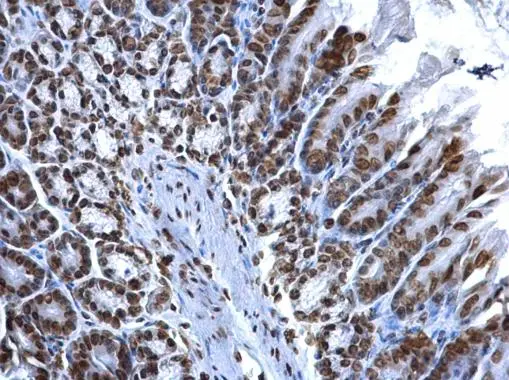
Histone H2A.X antibody detects Histone H2A.X protein at nucleus on mouse prostate by immunohistochemical analysis.
Sample: Paraffin-embedded mouse prostate.
Histone H2A.X antibody (GTX108272) dilution: 1:500.
Antigen Retrieval: Trilogy™ (EDTA based, pH 8.0) buffer, 15min
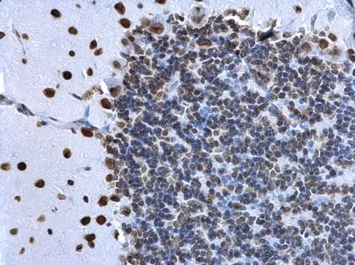
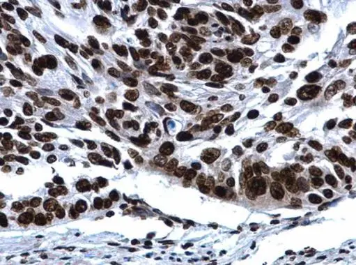
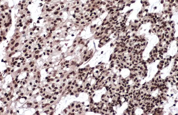
Histone H2A.X antibody detects Histone H2A.X protein at nucleus in human cervical cancer by immunohistochemical analysis.
Sample: Paraffin-embedded human cervical cancer.
Histone H2A.X antibody (GTX108272) diluted at 1:500.
Antigen Retrieval: Citrate buffer, pH 6.0, 15 min
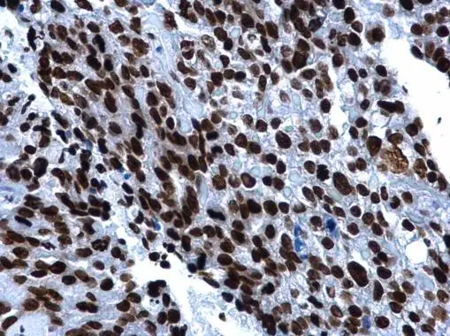
Histone H2A.X antibody detects Histone H2A.X protein at nucleus in human esophagus cancer by immunohistochemical analysis.
Sample: Paraffin-embedded human esophagus cancer.
Histone H2A.X antibody (GTX108272) diluted at 1:500.
Antigen Retrieval: Citrate buffer, pH 6.0, 15 min
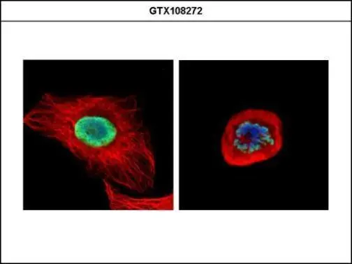
Confocal immunofluorescence analysis (Olympus FV10i) of paraformaldehyde-fixed U2OS, using Histone H2A.X (GTX108272) antibody (Green) at 1:500 dilution. Alpha-tubulin filaments were labeled with GTX11304 (Red) at 1:2000.
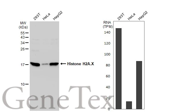
Various whole cell extracts (30 μg) were separated by 15% SDS-PAGE, and the membrane was blotted with Histone H2A.X antibody (GTX108272) diluted at 1:1000. The HRP-conjugated anti-rabbit IgG antibody (GTX213110-01) was used to detect the primary antibody. Corresponding RNA expression data for the same cell lines are based on Human Protein Atlas program.
Various whole cell extracts (30 μg) were separated by 15% SDS-PAGE, and the membrane was blotted with Histone H2A.X antibody (GTX108272) diluted at 1:500. The HRP-conjugated anti-rabbit IgG antibody (GTX213110-01) was used to detect the primary antibody.
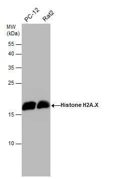
Various whole cell extracts (30 μg) were separated by 15% SDS-PAGE, and the membrane was blotted with Histone H2A.X antibody (GTX108272) diluted at 1:500. The HRP-conjugated anti-rabbit IgG antibody (GTX213110-01) was used to detect the primary antibody.
Histone H2A.X antibody detects Histone H2A.X protein by western blot analysis.
A. 50 μg mouse brain extract
12% SDS-PAGE
Histone H2A.X antibody (GTX108272) dilution: 1:1000
The HRP-conjugated anti-rabbit IgG antibody (GTX213110-01) was used to detect the primary antibody.
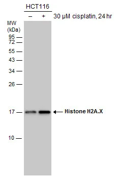
Untreated (–) and treated (+) HCT116 whole cell extracts (30 μg) were separated by 15% SDS-PAGE, and the membrane was blotted with Histone H2A.X antibody (GTX108272) diluted at 1:10000. The HRP-conjugated anti-rabbit IgG antibody (GTX213110-01) was used to detect the primary antibody.
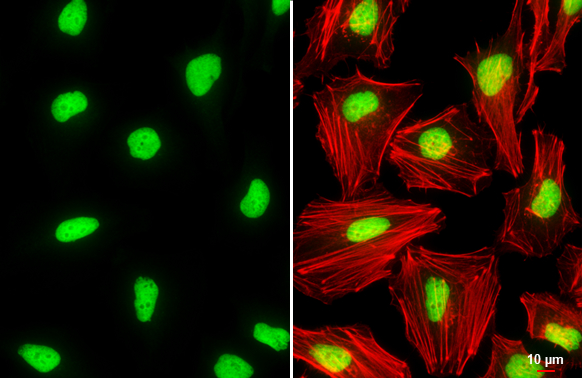
Histone H2A.X antibody detects Histone H2A.X protein at nucleus by immunofluorescent analysis.Sample: HeLa cells were fixed in 4% paraformaldehyde at RT for 15 min.Green: Histone H2A.X stained by Histone H2A.X antibody (GTX108272) diluted at 1:2000.Red: phalloidin, a cytoskeleton marker, diluted at 1:200.Scale bar= 10 μm.
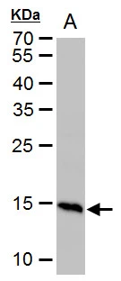
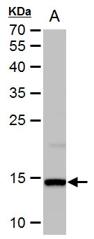
Histone H2A.X antibody detects Histone H2A.X protein by western blot analysis.
A. 50 μg rat brain extract
12% SDS-PAGE
Histone H2A.X antibody (GTX108272) dilution: 1:1000
The HRP-conjugated anti-rabbit IgG antibody (GTX213110-01) was used to detect the primary antibody.
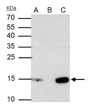
Histone H2A.X antibody immunoprecipitates H2AFX protein in IP experiments.
IP samples: Jurkat whole cell extract
A. 40 μg Jurkat whole cell extract
B. Control with 4 μg of preimmune Rabbit IgG
C. Immunoprecipitation of H2AFX protein by 4 μg Histone H2A.X antibody (GTX108272)
15 % SDS-PAGE
The immunoprecipitated H2AFX protein was detected by Histone H2A.X antibody (GTX108272) diluted at 1:2000.
[EasyBlot anti-rabbit IgG (GTX221666-01) was used as a secondary reagent]
Histone H2A.X antibody detects Histone H2A.X protein at nucleus by immunohistochemical analysis.Sample: Paraffin-embedded human breast carcinoma.Histone H2A.X stained by Histone H2A.X antibody (GTX108272) diluted at 1:500.Antigen Retrieval: Citrate buffer, pH 6.0, 15 min
-
HostRabbit
-
ClonalityPolyclonal
-
IsotypeIgG
-
ApplicationsWB ICC/IF IHC-P IP
-
ReactivityHuman, Mouse, Rat