Histone H2A.XS139ph (phospho Ser139) antibody


Untreated (–) and treated (+) HCT116 whole cell extracts (30 μg) were separated by 15% SDS-PAGE, and the membrane was blotted with Histone H2A.XS139ph (phospho Ser139) antibody (GTX127340) diluted at 1:1000. The HRP-conjugated anti-rabbit IgG antibody (GTX213110-01) was used to detect the primary antibody.
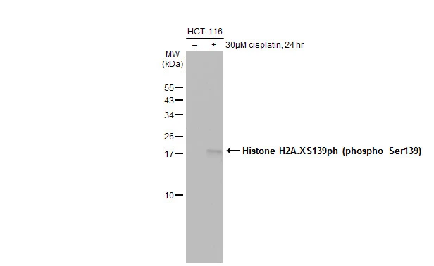
Untreated (–) and treated (+) HCT-116 whole cell extracts (30 μg) were separated by 15% SDS-PAGE, and the membrane was blotted with Histone H2A.XS139ph (phospho Ser139) antibody (GTX127340) diluted at 1:1000. The HRP-conjugated anti-rabbit IgG antibody (GTX213110-01) was used to detect the primary antibody.
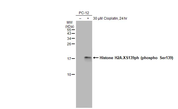
Untreated (–) and treated (+) PC-12 whole cell extracts (30 μg) were separated by 15% SDS-PAGE, and the membrane was blotted with Histone H2A.XS139ph (phospho Ser139) antibody (GTX127340) diluted at 1:1000. The HRP-conjugated anti-rabbit IgG antibody (GTX213110-01) was used to detect the primary antibody.
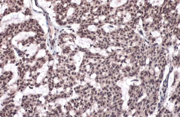
Histone H2A.XS139ph (phospho Ser139) antibody detects Histone H2A.XS139ph (phospho Ser139) protein at nucleus by immunohistochemical analysis.Sample: Paraffin-embedded human breast carcinoma.Histone H2A.XS139ph (phospho Ser139) stained by Histone H2A.XS139ph (phospho Ser139) antibody (GTX127340) diluted at 1:500.Antigen Retrieval: Citrate buffer, pH 6.0, 15 min
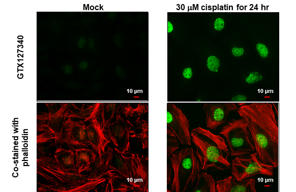
Histone H2A.XS139ph (phospho Ser139) antibody detects Histone H2A.XS139ph (phospho Ser139) protein at nucleus by immunofluorescent analysis.Sample: HeLa cells were fixed in 4% paraformaldehyde at RT for 15 min.Green: Histone H2A.XS139ph (phospho Ser139) stained by Histone H2A.XS139ph (phospho Ser139) antibody (GTX127340) diluted at 1:1000.Red: phalloidin, a cytoskeleton marker, diluted at 1:100.Scale bar= 10 μm.
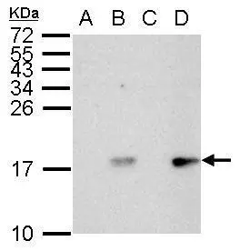
Histone H2A.X (phospho Ser139) antibody detects H2AFX protein by western blot analysis.
A. 30 μg HCT116 whole cell lysate/extract (untreated for 2hr)
B. 30 μg HCT116 whole cell lysate/extract (UVB treatment 50mJ for 2hr)
C. 30 μg HCT116 whole cell lysate/extract (untreated for 6hr)
D. 30 μg HCT116 whole cell lysate/extract (UVB treatment 50mJ for 6hr)
15% SDS-PAGE
Histone H2A.X (phospho Ser139) antibody (GTX127340) dilution: 1:1000
The HRP-conjugated anti-rabbit IgG antibody (GTX213110-01) was used to detect the primary antibody.
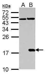
Histone H2A.X (phospho Ser139) antibody detects H2AFX protein in cisplatin-treated samples by western blot analysis.
A. 30 μg NIH-3T3 whole cell lysate/extract (untreated)
B. 30 μg NIH-3T3 whole cell lysate/extract (30μM cisplatin treatment for 24hr)
15% SDS-PAGE
Histone H2A.X (phospho Ser139) antibody (GTX127340) dilution: 1:1000
The HRP-conjugated anti-rabbit IgG antibody (GTX213110-01) was used to detect the primary antibody.
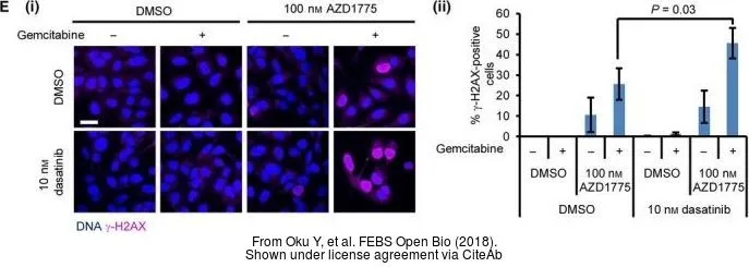
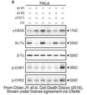
The data was published in the journal Cell Death Discov in 2016. PMID: 27551500
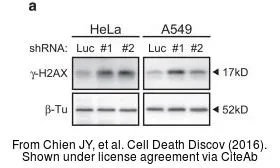
The data was published in the journal Cell Death Discov in 2016. PMID: 27551500
-
HostRabbit
-
ClonalityPolyclonal
-
IsotypeIgG
-
ApplicationsWB ICC/IF IHC-P IHC-Fr IHC-Wm
-
ReactivityHuman, Mouse, Rat, Zebrafish