Histone H3.3B antibody

Histone H3.3B antibody detects Histone H3.3B protein at nucleus on mouse spinal cord by immunohistochemical analysis.
Sample: Paraffin-embedded mouse spinal cord.
Histone H3.3B antibody (GTX115549) diluted at 1:500.
Antigen Retrieval: Trilogy™ (EDTA based, pH 8.0) buffer, 15min
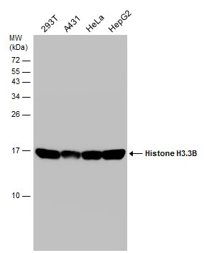
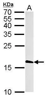
Various whole cell extracts (30 μg) were separated by 15% SDS-PAGE, and the membrane was blotted with Histone H3.3B antibody (GTX115549) diluted at 1:10000.
Sample (50 μg of whole cell lysate)
A: mouse brain
15% SDS PAGE
GTX115549 diluted at 1:1000
The HRP-conjugated anti-rabbit IgG antibody (GTX213110-01) was used to detect the primary antibody.
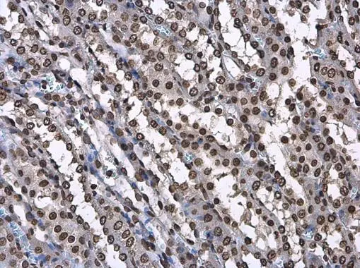
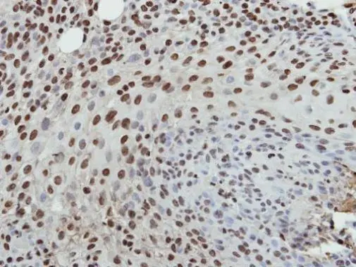
Histone H3.3B antibody detects Histone H3.3B protein at nucleus in rat kidney by immunohistochemical analysis.
Sample: Paraffin-embedded rat kidney.
Histone H3.3B antibody (GTX115549) diluted at 1:500.
Antigen Retrieval: Citrate buffer, pH 6.0, 15 min
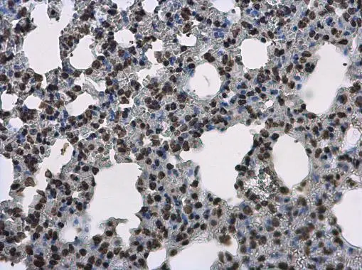
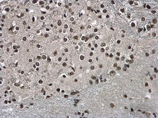
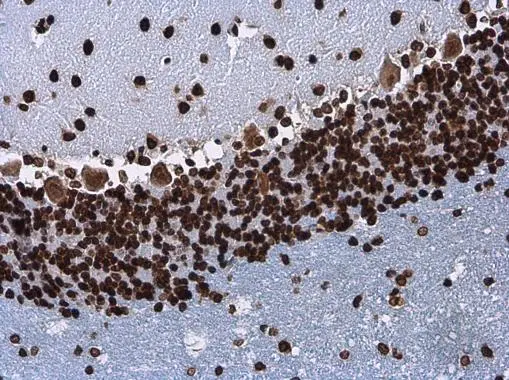
Histone H3.3B antibody detects Histone H3.3B protein at nucleus on mouse spinal cord by immunohistochemical analysis.
Sample: Paraffin-embedded mouse spinal cord.
Histone H3.3B antibody (GTX115549) diluted at 1:500.
Antigen Retrieval: Trilogy™ (EDTA based, pH 8.0) buffer, 15min
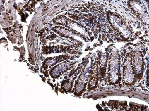
Histone H3.3B antibody detects Histone H3.3B protein at nucleus on mouse prostate by immunohistochemical analysis.
Sample: Paraffin-embedded mouse prostate.
Histone H3.3B antibody (GTX115549) dilution: 1:500.
Antigen Retrieval: Trilogy™ (EDTA based, pH 8.0) buffer, 15min
Histone H3.3B antibody detects Histone H3.3B protein at nucleus on mouse prostate by immunohistochemical analysis.
Sample: Paraffin-embedded mouse prostate.
Histone H3.3B antibody (GTX115549) dilution: 1:500.
Antigen Retrieval: Trilogy™ (EDTA based, pH 8.0) buffer, 15min
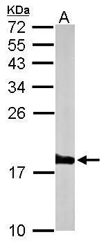
Histone H3.3B antibody detects H3F3B protein by western blot analysis.
A. 50 μg rat brain lysate/extract
15% SDS-PAGE
Histone H3.3B antibody (GTX115549) dilution: 1:1000
The HRP-conjugated anti-rabbit IgG antibody (GTX213110-01) was used to detect the primary antibody.
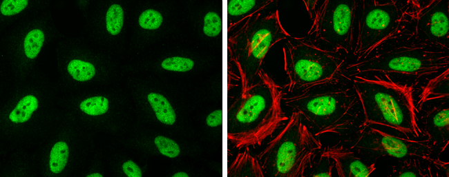
Histone H3.3B antibody detects Histone H3.3B protein at nucleus by immunofluorescent analysis.
Sample: HeLa cells were fixed in 4% paraformaldehyde at RT for 15 min.
Green: Histone H3.3B protein stained by Histone H3.3B antibody (GTX115549) diluted at 1:500.
Red: Phalloidin, a cytoskeleton marker, diluted at 1:100.
Immunohistochemical analysis of paraffin-embedded HSC3 xenograft, using Histone H3.3B(GTX115549) antibody at 1:500 dilution.
Antigen Retrieval: Trilogy™ (EDTA based, pH 8.0) buffer, 15min
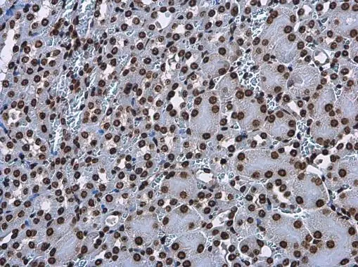
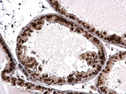
Histone H3.3B antibody detects Histone H3.3B protein at nucleus in mouse kidney by immunohistochemical analysis.
Sample: Paraffin-embedded mouse kidney.
Histone H3.3B antibody (GTX115549) diluted at 1:500.
Antigen Retrieval: Citrate buffer, pH 6.0, 15 min
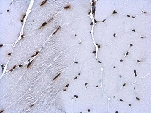
Histone H3.3B antibody detects Histone H3.3B protein at nucleus in mouse muscle by immunohistochemical analysis.
Sample: Paraffin-embedded mouse muscle.
Histone H3.3B antibody (GTX115549) diluted at 1:500.
Antigen Retrieval: Citrate buffer, pH 6.0, 15 min
Histone H3.3B antibody detects Histone H3.3B protein at nucleus in rat brain by immunohistochemical analysis.
Sample: Paraffin-embedded rat brain.
Histone H3.3B antibody (GTX115549) diluted at 1:500.
Antigen Retrieval: Citrate buffer, pH 6.0, 15 min
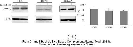
The data was published in the journal Evid Based Complement Alternat Med in 2013. PMID: 23533486
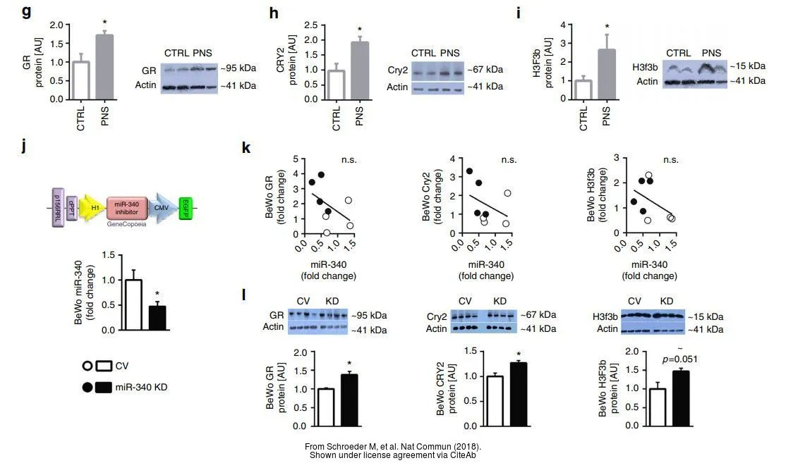
The data was published in the journal Nat Commun in 2018. PMID: 29686286
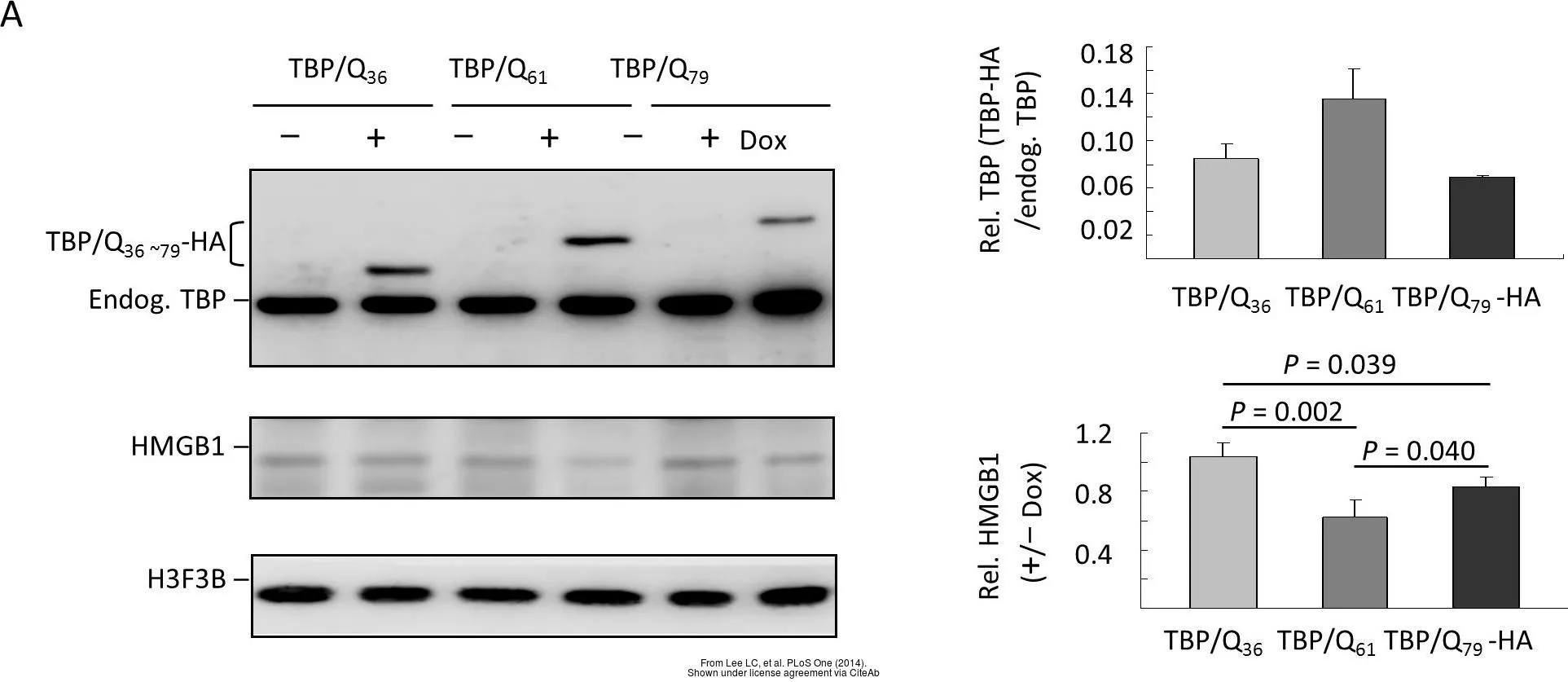
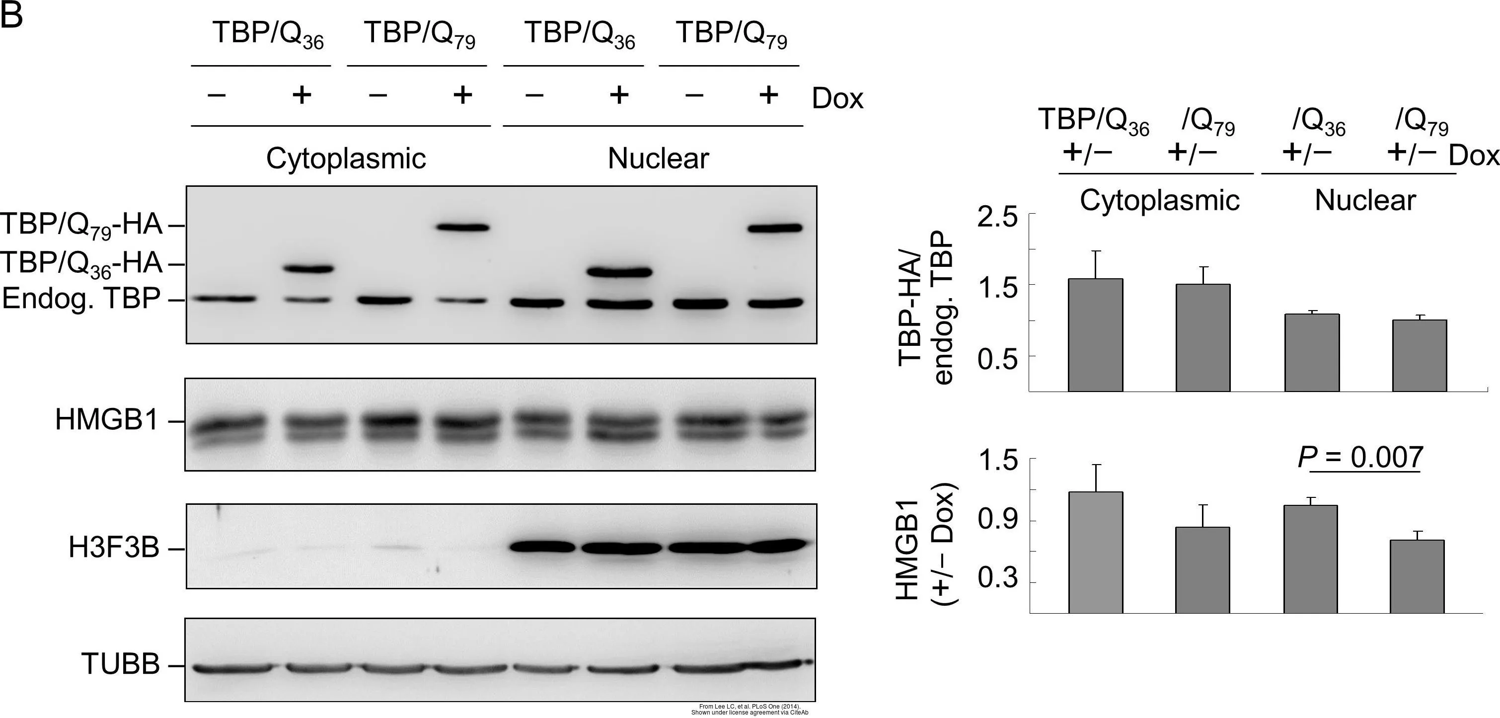
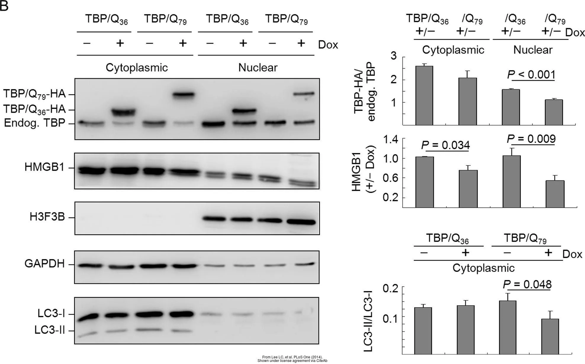
The data was published in the journal PLoS One in 2014.PMID: 25549101
-
HostRabbit
-
ClonalityPolyclonal
-
IsotypeIgG
-
ApplicationsWB ICC/IF IHC-P
-
ReactivityHuman, Mouse, Rat