Histone H3 antibody
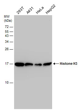
Various whole cell extracts (30 μg) were separated by 15% SDS-PAGE, and the membrane was blotted with Histone H3 antibody (GTX122150) diluted at 1:5000.
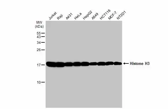
Various whole cell extracts (30 μg) were separated by 15% SDS-PAGE, and the membrane was blotted with Histone H3 antibody (GTX122150) diluted at 1:500. The HRP-conjugated anti-rabbit IgG antibody (GTX213110-01) was used to detect the primary antibody.
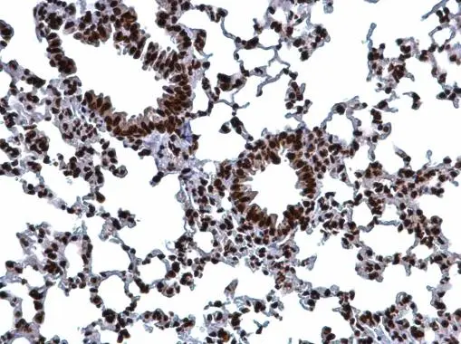
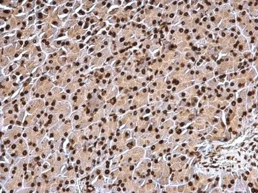
Histone H3.1 antibody detects Histone H3.1 protein at nucleus on mouse lung by immunohistochemical analysis.
Sample: Paraffin-embedded mouse lung.
Histone H3.1 antibody (GTX122150) dilution: 1:500.
Antigen Retrieval: Trilogy™ (EDTA based, pH 8.0) buffer, 15min
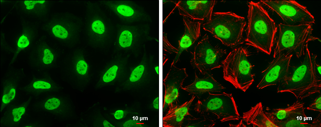
Histone H3 antibody detects Histone H3 protein at nucleus by immunofluorescent analysis.
Sample: HeLa cells were fixed in 4% paraformaldehyde at RT for 15 min.
Green: Histone H3 protein stained by Histone H3 antibody (GTX122150) diluted at 1:500.
Red: phalloidin, a cytoskeleton marker, diluted at 1:50.
Scale bar = 10 μm.
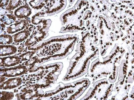
Histone H3.1 antibody detects Histone H3.1 protein at nucleus on mouse intestine by immunohistochemical analysis.
Sample: Paraffin-embedded mouse intestine.
Histone H3.1 antibody (GTX122150) dilution: 1:500.
Antigen Retrieval: Trilogy™ (EDTA based, pH 8.0) buffer, 15min
Histone H3.1 antibody detects Histone H3.1 protein at nucleus on mouse intestine by immunohistochemical analysis.
Sample: Paraffin-embedded mouse intestine.
Histone H3.1 antibody (GTX122150) dilution: 1:500.
Antigen Retrieval: Trilogy™ (EDTA based, pH 8.0) buffer, 15min
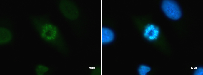
HIST1H3A antibody detects HIST1H3A protein at chromosome by immunofluorescent analysis.
Sample: HeLa cells were fixed in 4% paraformaldehyde/PBS for 15 min.
Green: HIST1H3A protein stained by HIST1H3A antibody (GTX122150) diluted at 1:500.
Blue: Hoechst 33342 staining.
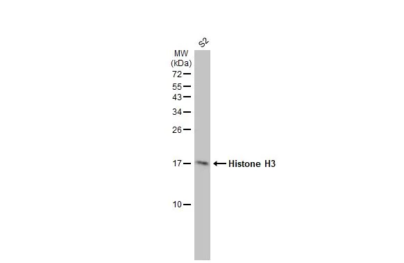
Whole cell extract (30 μg) was separated by 15% SDS-PAGE, and the membrane was blotted with Histone H3 antibody (GTX122150) diluted at 1:1000. The HRP-conjugated anti-rabbit IgG antibody (GTX213110-01) was used to detect the primary antibody.
-
HostRabbit
-
ClonalityPolyclonal
-
IsotypeIgG
-
ApplicationsWB ICC/IF IHC-P
-
ReactivityHuman, Mouse, Drosophila