Hsp70 antibody

Hsp70 antibody immunoprecipitates Hsp70 protein in IP experiments. IP Sample: 1000 μg HeLa whole cell lysate/extract A. 40 μg HeLa whole cell lysate/extract B. Control with 2.5 μg of preimmune rabbit IgG C. Immunoprecipitation of Hsp70 protein by 2.5 μg of Hsp70 antibody (GTX111088) 12% SDS-PAGE The immunoprecipitated Hsp70 protein was detected by Hsp70 antibody (GTX111088) diluted at 1:1000. EasyBlot anti-rabbit IgG (GTX221666-01) was used as a secondary reagent.
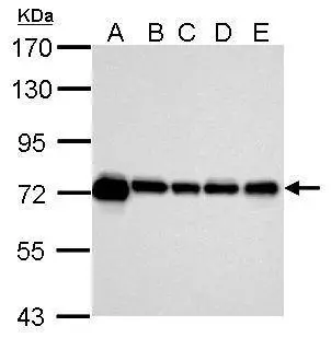
Sample (30 μg of whole cell lysate)
A: 293T
B: NIH-3T3
C: Mouse brain
D: PC-12
E: Rat brain
7.5% SDS PAGE
GTX111088 diluted at 1:10000
The HRP-conjugated anti-rabbit IgG antibody (GTX213110-01) was used to detect the primary antibody.
Various whole cell extracts were separated by 7.5% SDS-PAGE, and the membrane was blotted with Hsp70 antibody (GTX111088) diluted at 1:10000. The HRP-conjugated anti-rabbit IgG antibody (GTX213110-01) was used to detect the primary antibody.
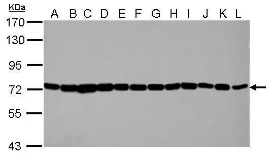
Sample (30 μg of whole cell lysate)
A: Jurkat
B: Raji
C: 293T
D: A431
E: HeLa
F: HepG2
G: H1299
H: HCT116
I: MCF-7
J: NT2D1
K: PC-3
L: U87-MG
7.5% SDS PAGE
GTX111088 diluted at 1:10000
The HRP-conjugated anti-rabbit IgG antibody (GTX213110-01) was used to detect the primary antibody.
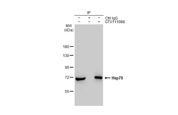
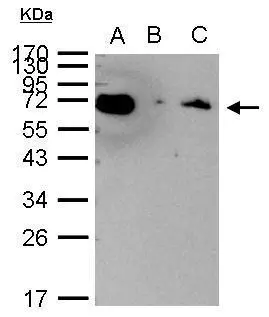
Immunoprecipitation of Hsp70 protein from HeLa whole cell extract using 5 μg of Hsp70 antibody (GTX111088).
Western blot analysis was performed using Hsp70 antibody (GTX111088).
EasyBlot HRP-conjugated anti rabbit IgG antibody (GTX221666-01).
Hsp70 antibody detects Hsp70 protein at cytosol on H1299 xenograft by immunohistochemical analysis.
Sample: Paraffin-embedded H1299 xenograft.
Hsp70 antibody (GTX111088) dilution: 1:500.
Antigen Retrieval: Trilogy™ (EDTA based, pH 8.0) buffer, 15min
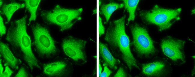
Hsp70 antibody detects Hsp70 protein at cytoplasm by immunofluorescent analysis.Sample: HeLa cells were fixed in ice-cold MeOH for 5 min.Green: Hsp70 stained by Hsp70 antibody (GTX111088) diluted at 1:500.Blue: Hoechst 33342 staining.
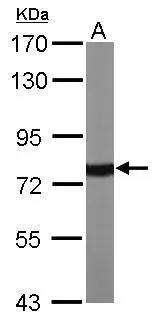
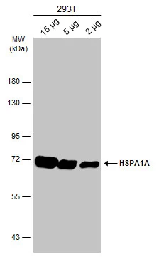
Sample (50 μg of whole cell lysate)
A: Mouse brain
7.5% SDS PAGE
GTX111088 diluted at 1:10000
The HRP-conjugated anti-rabbit IgG antibody (GTX213110-01) was used to detect the primary antibody.
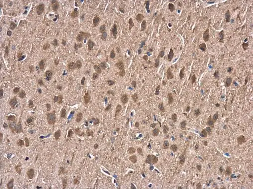
Hsp70 antibody detects Hsp70 protein at cytoplasm in rat brain by immunohistochemical analysis.
Sample: Paraffin-embedded rat brain.
Hsp70 antibody (GTX111088) diluted at 1:500.
Antigen Retrieval: Citrate buffer, pH 6.0, 15 min
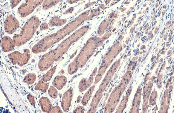
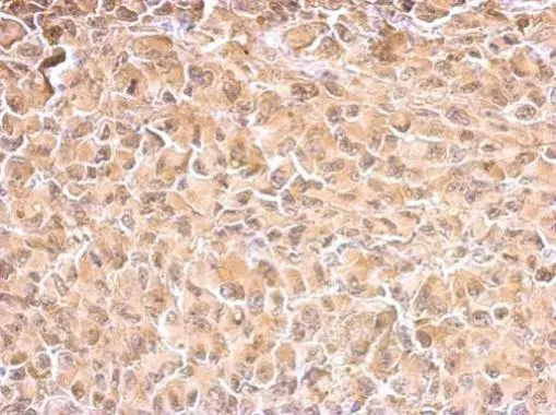
HSPA1A antibody detects HSPA1A protein at cytoplasm by immunohistochemical analysis.Sample: Paraffin-embedded human stomach.HSPA1A stained by HSPA1A antibody (GTX111088) diluted at 1:500.Antigen Retrieval: Citrate buffer, pH 6.0, 15 min
-
HostRabbit
-
ClonalityPolyclonal
-
IsotypeIgG
-
ApplicationsWB ICC/IF IHC-P IP
-
ReactivityHuman, Mouse, Rat, Monkey, Caenorhabditis elegans