APPLICATION
Application Note
*Optimal dilutions/concentrations should be determined by the researcher.
Application |
Recommended Dilution |
1:5000-1:20000 |
1:100-1:1000 |
1:100-1:1000 |
1:100-1:500 |
Not tested in other applications.
Calculated MW
Positive Control
mouse brain , rat brain , 293T , A431 , HeLa , HepG2
Product Note
Knockdown/Knockout validation was supported by customer review data.
Predict Reactivity
Bovine, Chicken, Pig, Chimpanzee, Rhesus Monkey(>80% identity)
PROPERTIES
Form
Liquid
Buffer
PBS, 1% BSA, 20% Glycerol
Preservative
0.025% ProClin 300
Storage
Store as concentrated solution. Centrifuge briefly prior to opening vial. For short-term storage (1-2 weeks), store at 4ºC. For long-term storage, aliquot and store at -20ºC or below. Avoid multiple freeze-thaw cycles.
Concentration
0.27 mg/ml (Please refer to the vial label for the specific concentration.)
Antigen Species
Human
Immunogen
Recombinant protein encompassing a sequence within the center region of human Hsp90 alpha. The exact sequence is proprietary.
Purification
Purified by antigen-affinity chromatography.
Conjugation
Unconjugated
RRID
AB_11178231
Note
For laboratory research use only. Not for any clinical, therapeutic, or diagnostic use in humans or animals. Not for animal or human consumption.
Purchasers shall not, and agree not to enable third parties to, analyze, copy, reverse engineer or otherwise attempt to determine the structure or sequence of the product.
TARGET
Synonyms
heat shock protein 90 alpha family class A member 1 , EL52 , HEL-S-65p , HSP86 , HSP89A , HSP90A , HSP90N , HSPC1 , HSPCA , HSPCAL1 , HSPCAL4 , HSPN , Hsp103 , Hsp89 , Hsp90 , LAP-2 , LAP2
Cellular Localization
Cytoplasm , Melanosome
Background
HSP90 proteins are highly conserved molecular chaperones that have key roles in signal transduction, protein folding, protein degradation, and morphologic evolution. HSP90 proteins normally associate with other cochaperones and play important roles in folding newly synthesized proteins or stabilizing and refolding denatured proteins after stress. There are 2 major cytosolic HSP90 proteins, HSP90AA1, an inducible form, and HSP90AB1 (MIM 140572), a constitutive form. Other HSP90 proteins are found in endoplasmic reticulum (HSP90B1; MIM 191175) and mitochondria (TRAP1; MIM 606219) (Chen et al., 2005 [PubMed 16269234]).[supplied by OMIM]
Database
Research Area
DATA IMAGES
|
GTX109753 WB Image
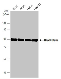
Various whole cell extracts (30 μg) were separated by 7.5% SDS-PAGE, and the membrane was blotted with Hsp90 alpha antibody (GTX109753) diluted at 1:10000. The HRP-conjugated anti-rabbit IgG antibody (GTX213110-01) was used to detect the primary antibody.
|
|
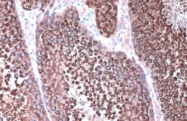
GTX109753 IHC-P Image
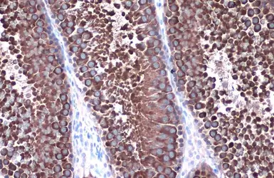
Hsp90 alpha antibody detects Hsp90 alpha protein at cytoplasm by immunohistochemical analysis.Sample: Paraffin-embedded rat testis.Hsp90 alpha stained by Hsp90 alpha antibody (GTX109753) diluted at 1:1000.Antigen Retrieval: Citrate buffer, pH 6.0, 15 min
|
|
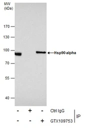
GTX109753 IP Image
Immunoprecipitation of Hsp90 alpha protein from 293T whole cell extracts using 5 μg of Hsp90 alpha antibody (GTX109753). Western blot analysis was performed using Hsp90 alpha antibody (GTX109753). EasyBlot anti-Rabbit IgG (GTX221666-01) was used as a secondary reagent.
|
|
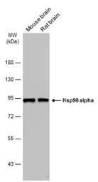
GTX109753 WB Image
Various tissue extracts (50 μg) were separated by 7.5% SDS-PAGE, and the membrane was blotted with Hsp90 alpha antibody (GTX109753) diluted at 1:10000. The HRP-conjugated anti-rabbit IgG antibody (GTX213110-01) was used to detect the primary antibody.
|
|
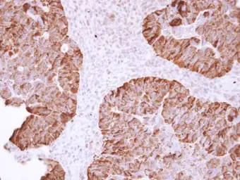
GTX109753 IHC-P Image
Hsp90 alpha antibody detects Hsp90 alpha protein at cytoplasm in human colon carcinoma by immunohistochemical analysis. Sample: Paraffin-embedded human colon carcinoma. Hsp90 alpha antibody (GTX109753) diluted at 1:500.
Antigen Retrieval: Citrate buffer, pH 6.0, 15 min
|
|
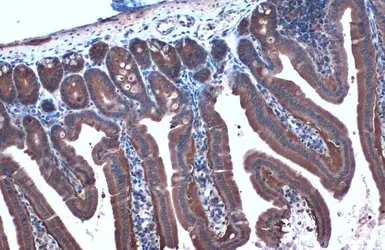
GTX109753 IHC-P Image
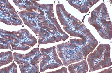
Hsp90 alpha antibody detects Hsp90 alpha protein at cytoplasm by immunohistochemical analysis.Sample: Paraffin-embedded mouse intestine.Hsp90 alpha stained by Hsp90 alpha antibody (GTX109753) diluted at 1:1000.Antigen Retrieval: Citrate buffer, pH 6.0, 15 min
|
|
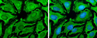
GTX109753 ICC/IF Image
Hsp90 alpha antibody detects Hsp90 alpha protein at cytoplasm by immunofluorescent analysis.Sample: HeLa cells were fixed in ice-cold MeOH for 5 min.Green: Hsp90 alpha stained by Hsp90 alpha antibody (GTX109753) diluted at 1:500.Blue: Hoechst 33342 staining.
|
|
GTX109753 IHC-P Image
Hsp90 alpha antibody detects Hsp90 alpha protein at cytoplasm by immunohistochemical analysis.Sample: Paraffin-embedded rat testis.Hsp90 alpha stained by Hsp90 alpha antibody (GTX109753) diluted at 1:1000.Antigen Retrieval: Citrate buffer, pH 6.0, 15 min
|
|
GTX109753 IHC-P Image
Hsp90 alpha antibody detects Hsp90 alpha protein at cytoplasm by immunohistochemical analysis.Sample: Paraffin-embedded mouse duodenum.Hsp90 alpha stained by Hsp90 alpha antibody (GTX109753) diluted at 1:1000.Antigen Retrieval: Citrate buffer, pH 6.0, 15 min
|
|
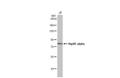
GTX109753 WB Image
Whole cell extract (30 μg) was separated by 7.5% SDS-PAGE, and the membrane was blotted with Hsp90 alpha antibody (GTX109753) diluted at 1:1000. The HRP-conjugated anti-rabbit IgG antibody (GTX213110-01) was used to detect the primary antibody.
Drosophila Hsp83 is the homolog of mammalian Hsp90. (Gene ID: 38389/ UniProt: P02828)
|
|
GTX109753 WB Image
The data was published in the journal Cells in 2019. PMID: 31905895
|
|
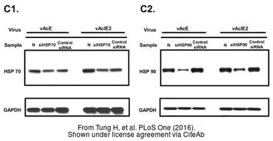
GTX109753 WB Image
The data was published in the journal PLoS One in 2016. PMID: 26863132
|
|
GTX109753 WB Image
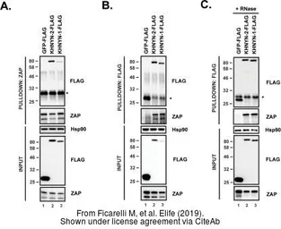
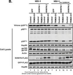
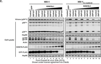
The data was published in the journal Elife in 2019.PMID: 31284899
|
|
GTX109753 WB Image
The data was published in the journal Elife in 2019.PMID: 31284899
|
|
GTX109753 WB Image
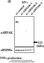
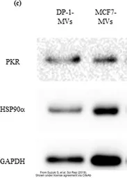
The data was published in the journal Sci Rep in 2019.PMID: 30846715
|
|
GTX109753 WB Image
The data was published in the journal Elife in 2019.PMID: 31284899
|
|
GTX109753 WB Image
The data was published in the journal Sci Rep in 2019.PMID: 30846715
|
REFERENCE
REVIEW
Hsp90 alpha antibody
Cat. No. GTX109753
Rating
|
|
( Average 5 based on 4 users reviews)
|
Application
Western Blot(WB)
|
|
( Average 5 based on 3 users reviews)
|
Immunoprecipitation(IP)
|
|
( Average 5 based on 1 users reviews)
|
Date :
Anonymous submitted on 02-Mar-2018
Application Tested :
WB
Sample Species :
Hu
Sample :
PC-3 cells
Blocking :
5% ECL Blocking Agent, 25°C, 1Hr
Primary Antibody :
1:5000, 4°C, 16Hr
Lane Description :
Lane 1: WCL 10 μg, Lane 2: heat shock (43oC 1.5h) WCL 4 μg, Lane 3: heat shock (43oC 3.0h) WCL 8 μg
Date :
Anonymous submitted on 26-Jul-2017
Application Tested :
WB
Sample Species :
Ms
Sample :
MC3T3-E1 cells
Amount used :
50 μg
Blocking :
5% milk in TBST, 25°C, 0.5Hr
Primary Antibody :
1:2000, 4°C, 18Hr
Date :
Anonymous submitted on 26-Apr-2017
Application Tested :
WB
Sample Species :
Hu
Sample :
HT29 cells
Blocking :
5% skim milk in PBST, 25°C, 1Hr
Primary Antibody :
1:5000, 4°C, 16Hr
Lane Description :
Lane1: HT29 shLacZ (virus infection to silence LacZ gene), Lane2: HT29 shHSP90AA1 ( virus infection to silence HSP90AA1 gene)
Date :
Anonymous submitted on 18-Jan-2013
Application Tested :
IP
Sample Species :
Rat
Sample :
RK3E cells
Amount used :
50μg
Blocking :
5% non-fat milk, 25°C, 1Hr
Primary Antibody :
1:5000, 4°C, 16Hr
Figure Note :
2ug for IP