ID1 antibody


*The competitor is not affiliated with GeneTex and does not endorse this product.
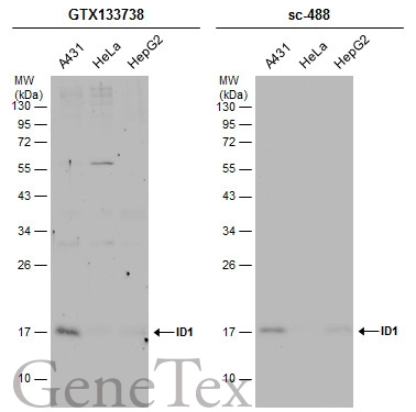
Various whole cell extracts (30 μg) were separated by 12% SDS-PAGE, and the membranes were blotted with ID1 antibody (GTX133738) diluted at 1:500 and competitor's antibody (sc-488) diluted at 1:500. The HRP-conjugated anti-rabbit IgG antibody (GTX213110-01) was used to detect the primary antibody.

Mouse whole cell extract (50 μg) was separated by 15% SDS-PAGE, and the membrane was blotted with ID1 antibody (GTX133738) diluted at 1:500. The HRP-conjugated anti-rabbit IgG antibody (GTX213110-01) was used to detect the primary antibody.
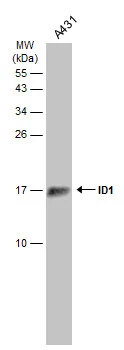
Whole cell extract (30 μg) was separated by 15% SDS-PAGE, and the membrane was blotted with ID1 antibody (GTX133738) diluted at 1:500. The HRP-conjugated anti-rabbit IgG antibody (GTX213110-01) was used to detect the primary antibody.
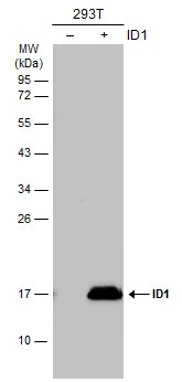
Non-transfected (–) and transfected (+) 293T whole cell extracts (30 μg) were separated by 12% SDS-PAGE, and the membrane was blotted with ID1 antibody (GTX133738) diluted at 1:1000. The HRP-conjugated anti-rabbit IgG antibody (GTX213110-01) was used to detect the primary antibody.
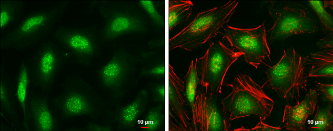
ID1 antibody detects ID1 protein at cytoplasm and nucleus by immunofluorescent analysis.Sample: HeLa cells were fixed in 4% paraformaldehyde at RT for 15 min.Green: ID1 stained by ID1 antibody (GTX133738) diluted at 1:1000.Red: phalloidin, a cytoskeleton marker, diluted at 1:100.Scale bar= 10μm.
-
HostRabbit
-
ClonalityPolyclonal
-
IsotypeIgG
-
ApplicationsWB ICC/IF
-
ReactivityHuman, Mouse