GeneTex
  • Country / Location Selection

United States (US)

IL27 antibody

Anti-IL27 antibody used in Western Blot (WB). GTX48686

Cat. No. GTX48686

Host

Rabbit

Clonality

Polyclonal

Isotype

IgG

Application

WB, ELISA

Reactivity

Mouse
Package
50 μg ($399)

APPLICATION

Application Note

*Optimal dilutions/concentrations should be determined by the researcher.
Application Recommended Dilution
WB 1:500-1:2000
ELISA 1:1000-1:5000
Not tested in other applications.

Calculated MW

27 kDa. ( Note )

PROPERTIES

Form

Liquid

Buffer

20mM Potassium Phosphate, 150mM NaCl

Preservative

No preservative

Storage

Store as concentrated solution. Centrifuge briefly prior to opening vial. For short-term storage (1-2 weeks), store at 4ºC. For long-term storage, aliquot and store at -20ºC or below. Avoid multiple freeze-thaw cycles.

Concentration

1 mg/ml (Please refer to the vial label for the specific concentration.)

Antigen Species

Mouse

Immunogen

Full length recombinant mouse IL27/p28 protein.

Purification

IgG fraction
This product is an IgG fraction antibody purified from monospecific antiserum by a multi-step process which includes delipidation, salt fractionation and ion exchange chromatography followed by extensive dialysis against the buffer stated above.

Conjugation

Unconjugated

Note

For laboratory research use only. Not for any clinical, therapeutic, or diagnostic use in humans or animals. Not for animal or human consumption.

Purchasers shall not, and agree not to enable third parties to, analyze, copy, reverse engineer or otherwise attempt to determine the structure or sequence of the product.

TARGET

Synonyms

interleukin 27 , IL-27 , IL-27-A , IL-27p28 , IL27-A , Il30 , p28

Cellular Localization

Secreted

Background

The cytokine Interleukin 27 (IL-27) is produced in response to inflammation. It is made by activated antigen presenting cells including monocytes, endothelial cells, and dendritic cells. IL-27 consists of a heterodimeric combination of Epstein-Barr virus-induced molecule 3 (EBI3, or IL-27B) non-covalently linked with IL-27 p28 (or IL-27A). It is a regulator of T helper cell development and suppressor of T-cell proliferation. IL-27 has both pro- and anti-inflammatory properties. It can stimulate cytotoxic T cell activity and induce isotype switching in B-cells. It has diverse effects on innate immune cells. It induces monocytes and mast cells to secrete pro-inflammatory cytokines. When infection is present, IL-27 induces naive CD4+ T cells to proliferate and develop Th1 cell responses. As an anti-inflammatory regulator, IL-27 can inhibit Th1 or Th2 responses and restrict the strength and duration of adaptive immune responses. The IL-27 p28 subunit, a 28 kDa glycoprotein belonging to the type I cytokine family, is homologous to IL-12 p35, IL-23 p19, and IL-6. The EBI3 (Epstein-Barr virus-induced molecule 3, or IL-27B) subunit is a 23.6 kDa glycoprotein containing two fibronectin type III domains, and belongs to the type I cytokine receptor family. It can exist as a homodimer and can also heterodimerize with IL-12 p35. It is homologous to the p40 subunit of IL-12 and IL-23 and to the extracellular domain of IL-6 R. EBI3 can heterodimerize also with IL-12 p35, or can exist as a homodimer. The heterodimeric IL-27 receptor contains WSX-1 (TCCR) and gp130. WSX-1 is specific for IL-27, and is expressed on resting/naive CD4+ T cells, CD8+ T cells, NK cells, dendritic cells, monocytes, mast cells, and B cells. Gp130, on the other hand, functions as a subunit of the receptor complexes for at least seven other cytokines. IL-27 also promotes effector functions of NK cells and CD8+ T cells.

Database

Research Area

DATA IMAGES

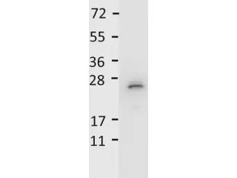
Anti-IL27 antibody used in Western Blot (WB). GTX48686

GTX48686 WB Image

Detection of recombinant IL27/p28 protein by anti-Mouse IL-27/p28 antibody (GTX48686). Recombinant mouse IL27/p28 was loaded on to an SDS-PAGE gel at 0.25 μg and after separation, transferred to nitrocellulose. The membrane was blocked with 1% BSA in TBST for 30 min at RT, followed by incubation with primary antibody diluted 1:1,000 in 1% BSA in TBST overnight at 4ºC. After washes, the blot was reacted with secondary antibody HRP Goat anti-Rabbit IgG antibody diluted 1:40,000 in blocking buffer for 30 min at RT. Data was collected using Bio-Rad VersaDocR 4000 MP imaging system.

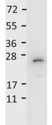
Anti-IL27 antibody used in Western Blot (WB). GTX48686

GTX48686 WB Image

WB analysis of recombinant mouse IL27 protein using GTX48686 IL27 antibody.
Loading : 0.25 μg
Dilution : 1:1000

REFERENCE

There are currently no references for IL27 antibody (GTX48686). Be the first to share your publications with this product.

REVIEW

There are currently no reviews for IL27 antibody (GTX48686). Be the first to share your experience with this product.
Package List Price ($)
$ 399