Iaspp antibody
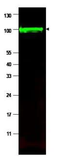
WB analysis of MCF-7 whole cell lysate using GTX48696 Iaspp antibody.
Loading : 35 μg
Dilution : 1:500
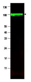
Western blot using GeneTex's affinity purified anti-iASPP antibody shows detection of a band at ~100 kDa (arrowhead) corresponding to isoform 1 of iASPP in MCF7 whole cell lysates. Preincubation with immunizing peptide blocks specific band staining (data not shown). Approximately 35 μg of lysate was separated by 4-20% Tris Glycine SDS-PAGE. After blocking, the membrane was probed with the primary antibody diluted to 1:1,500 in 5% BLOTTO/PBS overnight at 4ºC. The membrane was washed and reacted with a 1:10,000 dilution of IRDye800 conjugated goat anti-Rabbit IgG [H&L] for 45 min at room temperature (800 nm channel, green). Molecular weight estimation was made by comparison to prestained MW markers. IRDye800 fluorescence image was captured using the OdysseyR Infrared Imaging System developed by LI-COR. IRDye is a trademark of LI-COR, Inc. Other detection systems will yield similar results.
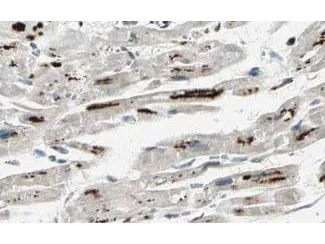
GeneTex's Affinity Purified anti-iASPP antibody shows strong cytoplasmic and membranous staining of myocytes in human heart tissue. Tissue was formalin-fixed and paraffin embedded. Brown color indicates presence of protein, blue color shows cell nuclei.
-
HostRabbit
-
ClonalityPolyclonal
-
IsotypeIgG
-
ApplicationsWB IHC-P ELISA
-
ReactivityHuman