 GeneTex
Iba1 antibody


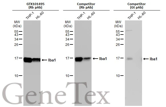
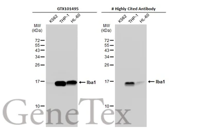
Various whole cell extracts (30 μg) were separated by 15% SDS-PAGE, and the membranes were blotted with Iba1 antibody (GTX101495) diluted at 1:1000 and competitor's antibody diluted at 1:1000. The HRP-conjugated anti-rabbit IgG antibody (GTX213110-01) was used to detect the primary antibody.
*The competitor is not affiliated with GeneTex and does not endorse this product.
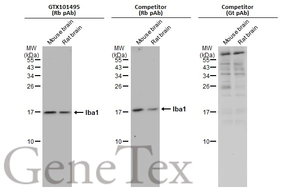
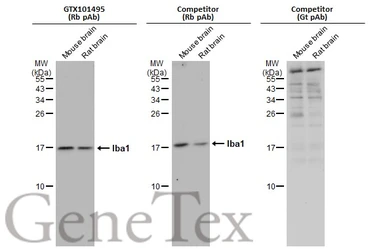
Various tissue extracts (50 μg) were separated by 15% SDS-PAGE, and the membranes were blotted with Iba1 antibody (GTX101495) diluted at 1:1000 and competitor's antibody diluted at 1:1000. The HRP-conjugated anti-rabbit IgG antibody (GTX213110-01) was used to detect the primary antibody, and the signal was developed with Trident ECL plus-Enhanced.
*The competitor is not affiliated with GeneTex and does not endorse this product.
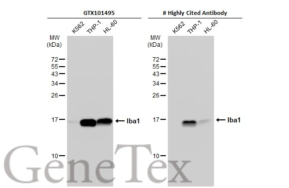
Various whole cell extracts (30 μg) were separated by 15% SDS-PAGE, and the membrane was blotted with Iba1 antibody (GTX101495) diluted at 1:5000. The HRP-conjugated anti-rabbit IgG antibody (GTX213110-01) was used to detect the primary antibody.
*The competitor is not affiliated with GeneTex and does not endorse this product.
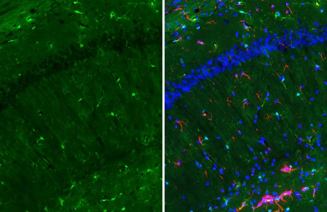
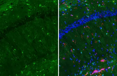
Iba1 antibody detects Iba1 protein by immunohistochemical analysis.Sample: Frozen-sectioned mouse hippocampus.Green: Iba1 stained by Iba1 antibody (GTX101495) diluted at 1:1000.Red: GFAP antibody [GT25] (GTX641104) diluted at 1:750.Blue: Fluoroshield with DAPI (GTX30920).
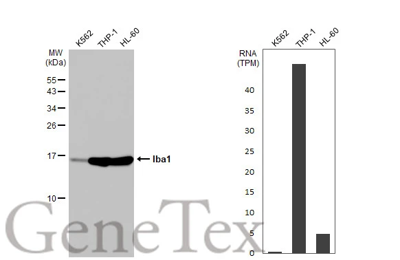
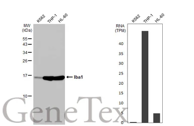
Various whole cell extracts (30 μg) were separated by 15% SDS-PAGE, and the membrane was blotted with Iba1 antibody (GTX101495) diluted at 1:5000. The HRP-conjugated anti-rabbit IgG antibody (GTX213110-01) was used to detect the primary antibody. Corresponding RNA expression data for the same cell lines are based on Human Protein Atlas program.
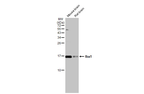
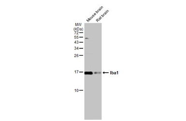
Various tissue extracts (50 μg) were separated by 15% SDS-PAGE, and the membrane was blotted with Iba1 antibody (GTX101495) diluted at 1:500. The HRP-conjugated anti-rabbit IgG antibody (GTX213110-01) was used to detect the primary antibody.
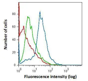
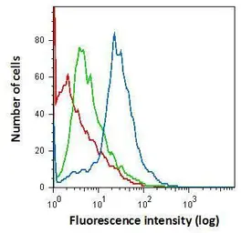
Flow cytometry on primary murine microglia cells, staining with Iba1 (GTX101495) antibody using 1.0 μg per 4×10⁵ cells. GTX101495 (blue), Rabbit IgG (green) ,Unstained (red).

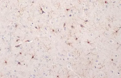
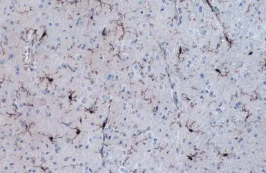
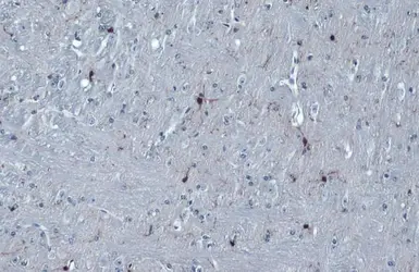
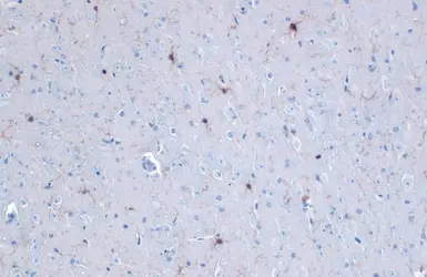
Iba1 antibody detects Iba1 protein at cell membrane and cytoplasm by immunohistochemical analysis.
Sample: Paraffin-embedded rat brain.
Iba1 stained by Iba1 antibody (GTX101495) diluted at 1:500.
Antigen Retrieval: Citrate buffer, pH 6.0, 15 min
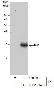
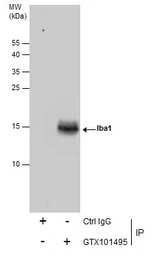
Immunoprecipitation of Iba1 protein from K562 whole cell extracts using 5 μg of Iba1 antibody (GTX101495).
Western blot analysis was performed using Iba1 antibody (GTX101495).
EasyBlot anti-Rabbit IgG (GTX221666-01) was used as a secondary reagent.
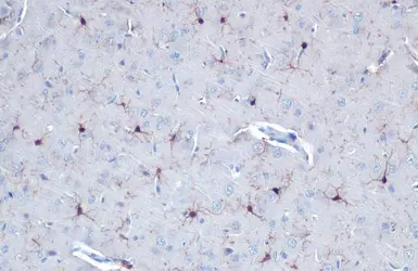
Iba1 antibody detects Iba1 protein at cell membrane and cytoplasm by immunohistochemical analysis.
Sample: Paraffin-embedded mouse brain.
Iba1 stained by Iba1 antibody (GTX101495) diluted at 1:500.
Antigen Retrieval: Citrate buffer, pH 6.0, 15 min
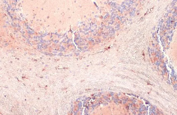
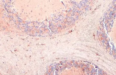
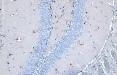
Iba1 antibody detects Iba1 protein at cell membrane and cytoplasm by immunohistochemical analysis.Sample: Paraffin-embedded rat cerebellum.Iba1 stained by Iba1 antibody (GTX101495) diluted at 1:1000.Antigen Retrieval: Citrate buffer, pH 6.0, 15 min
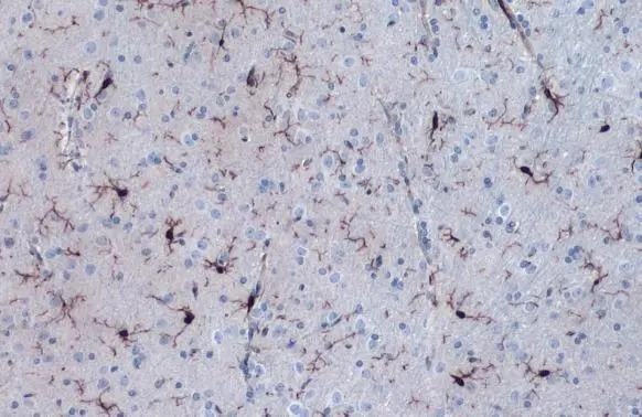
Iba1 antibody detects Iba1 protein at cell membrane and cytoplasm by immunohistochemical analysis.Sample: Paraffin-embedded mouse cerebellum.Iba1 stained by Iba1 antibody (GTX101495) diluted at 1:1000.Antigen Retrieval: Citrate buffer, pH 6.0, 15 min
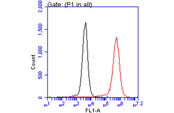
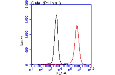
Iba1 antibody (GTX101495) detects AIF1 protein by flow cytometry analysis.
Sample: THP-1 cell.
Black: Unlabelled sample was used as a control.
Red: Iba1 antibody (GTX101495) dilution: 1:50.
Acquisition of 20,000 events were collected for FACS analysis.
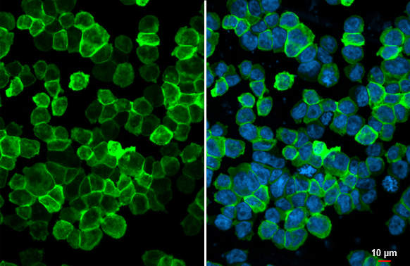
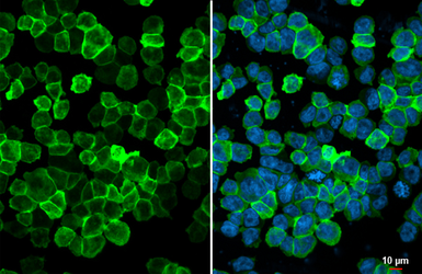
Iba1 antibody detects Iba1 protein at cell periphery by immunofluorescent analysis.Sample: THP-1 cells were fixed in 4% paraformaldehyde at RT for 15 min.Green: Iba1 stained by Iba1 antibody (GTX101495) diluted at 1:500.Blue: Fluoroshield with DAPI (GTX30920).
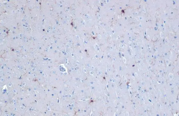
Iba1 antibody detects Iba1 protein at cell membrane and cytoplasm by immunohistochemical analysis.Sample: Paraffin-embedded mouse brain.Iba1 stained by Iba1 antibody (GTX101495) diluted at 1:500.Antigen Retrieval: Citrate buffer, pH 6.0, 15 min
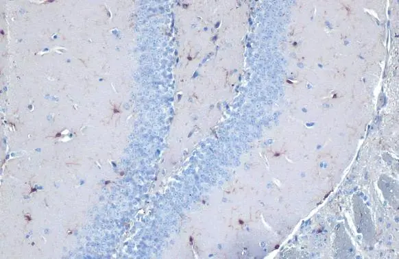
Iba1 antibody detects Iba1 protein at cell membrane and cytoplasm by immunohistochemical analysis.Sample: Paraffin-embedded mouse brain.Iba1 stained by Iba1 antibody (GTX101495) diluted at 1:500.Antigen Retrieval: Citrate buffer, pH 6.0, 15 min
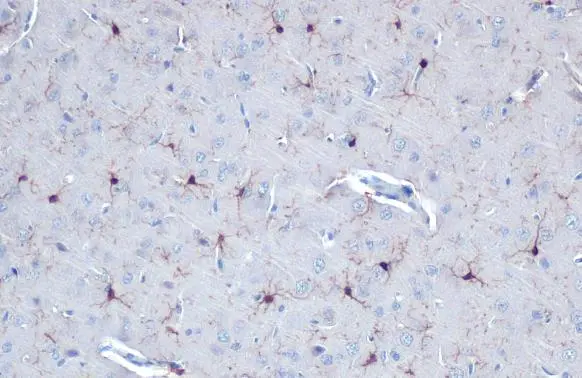
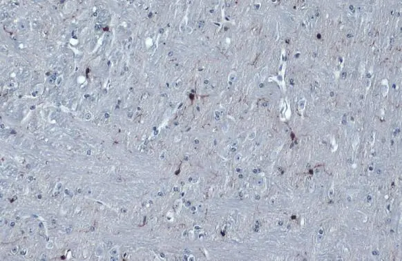
Iba1 antibody detects Iba1 protein at cell membrane and cytoplasm by immunohistochemical analysis.Sample: Paraffin-embedded rat brain.Iba1 stained by Iba1 antibody (GTX101495) diluted at 1:500.Antigen Retrieval: Citrate buffer, pH 6.0, 15 min

-
HostRabbit
-
ClonalityPolyclonal
-
IsotypeIgG
-
ApplicationsWB ICC/IF IHC-P IHC-Fr FCM IP
-
ReactivityHuman, Mouse, Rat