KCNK1 antibody
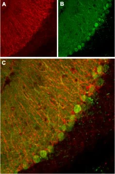
IHC-Fr analysis of mouse cerebellum tissue using GTX16685 KCNK1 antibody.
Panel A : KCNK1 channel appears in glial processes (red).
Panel B : Staining of Purkinje nerve cells with mouse anti-calbindin D28K (a calcium binding protein, green).
Panel C : Merge of KCNK1 channel and calbindin D28K demonstrates the separate localization of these proteins.
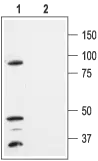
WB analysis of HEK-293-KCNK1 transfected cell lysates using GTX16685 KCNK1 antibody preincubated with or without immunogen peptide.
Dilution : 1:300
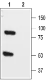
WB analysis of rat brain lysate using GTX16685 KCNK1 antibody preincubated with or without immunogen peptide.
Dilution : 1:200
-
HostRabbit
-
ClonalityPolyclonal
-
IsotypeIgG
-
ApplicationsWB IHC-Fr
-
ReactivityHuman, Mouse, Rat