KIR2DS2 antibody
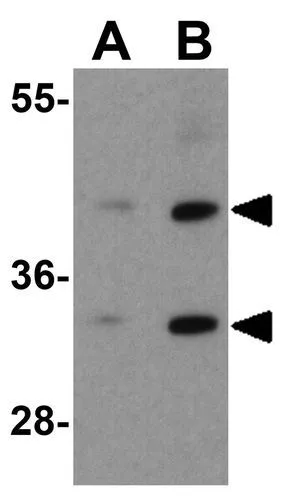
WB analysis of 293 cell lysate using GTX31949 KIR2DS2 antibody.
Working concentration : (A) 1 and (B) 2 μg/ml
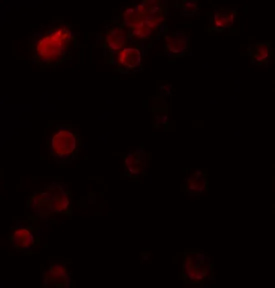
ICC/IF analysis of HEK293 cells using GTX31949 KIR2DS2 antibody.
Dilution : 5 μg/ml
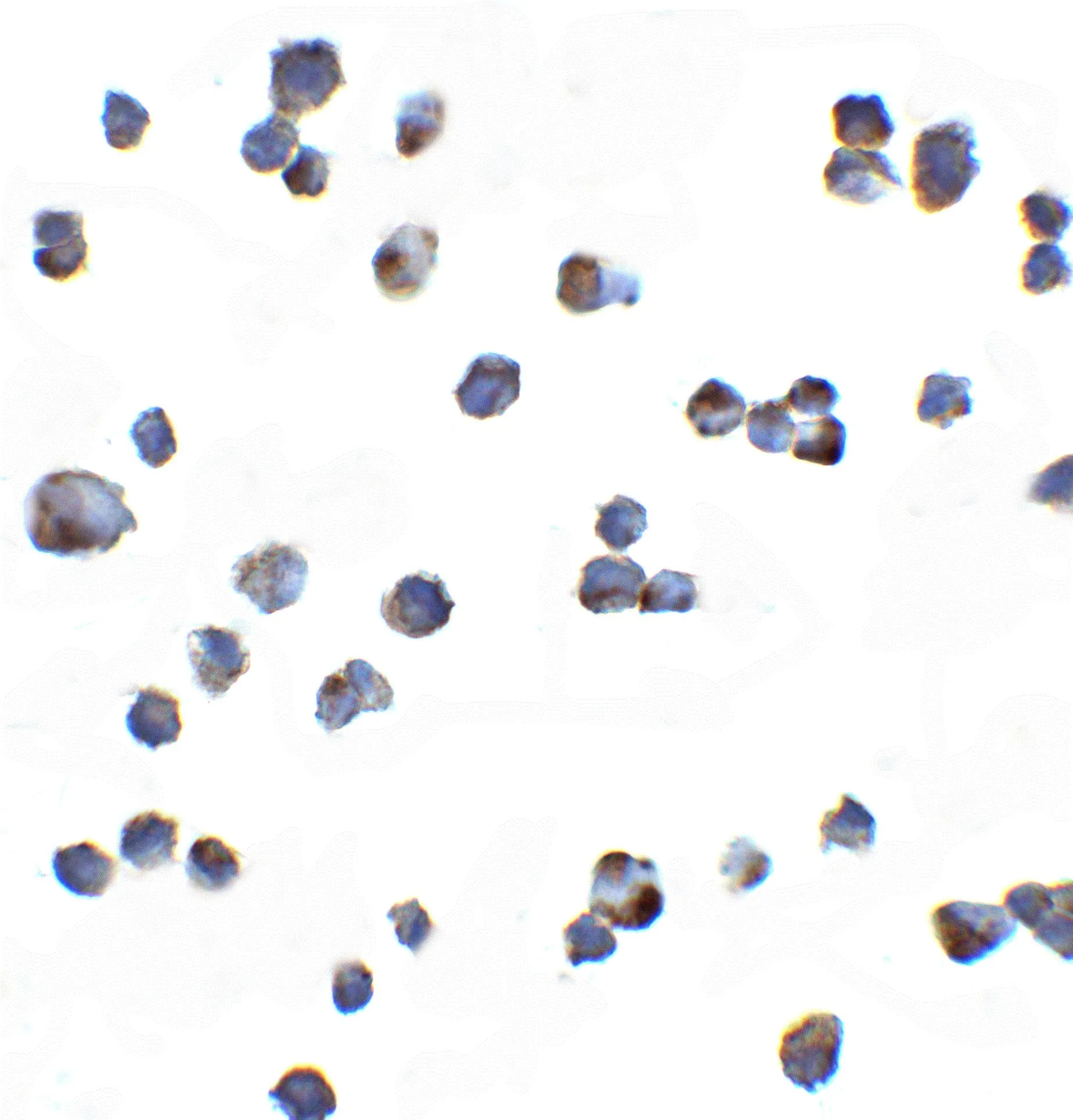
ICC/IF analysis of 293 cells using GTX31949 KIR2DS2 antibody.
Working concentration : 20 μg/ml
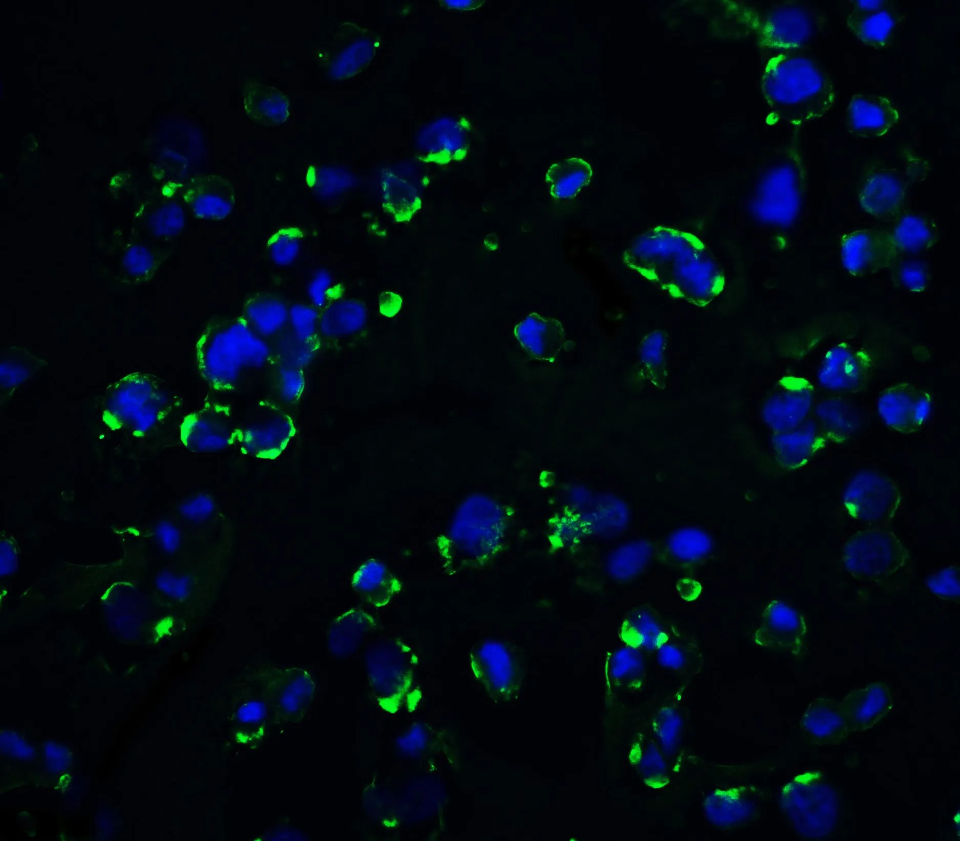
ICC/IF analysis of HEK293 cells using GTX31949 KIR2DS2 antibody.
Green : Primary antibody
Blue : DAPI
Dilution : 20 μg/ml
-
HostRabbit
-
ClonalityPolyclonal
-
IsotypeIgG
-
ApplicationsWB ICC/IF ELISA
-
ReactivityHuman