L-Plastin antibody
Cat. No. GTX33300
Cat. No. GTX33300
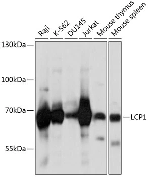
GTX33300 WB Image
WB analysis of various sample lysates using GTX33300 L-Plastin antibody.
Dilution : 1:1000
Loading : 25μg per lane
1 / 2
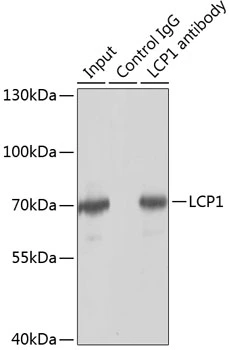
GTX33300 IP Image
IP analysis of Jurkat cell lysate using GTX33300 L-Plastin antibody.
Antibody amount : 3μg / 150μg lysate
Dilution : 1:1000
2 / 2
-
HostRabbit
-
ClonalityPolyclonal
-
IsotypeIgG
-
ApplicationsWB IP
-
ReactivityHuman, Mouse