LAMP2 antibody


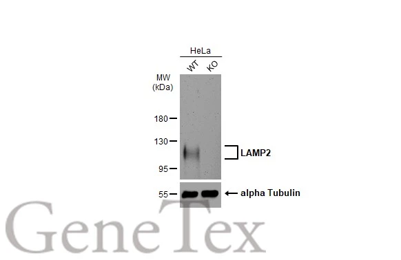
Wild-type (WT) and LAMP2 knockout (KO) HeLa cell extracts (30 μg) were separated by 7.5% SDS-PAGE, and the membrane was blotted with LAMP2 antibody (GTX103214) diluted at 1:5000. The HRP-conjugated anti-rabbit IgG antibody (GTX213110-01) was used to detect the primary antibody, and the signal was developed with Trident ECL plus-Enhanced.
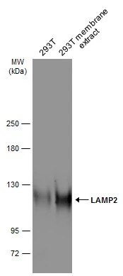
293T whole cell and membrane extracts (30 μg) were separated by 5% SDS-PAGE, and the membrane was blotted with LAMP2 antibody (GTX103214) diluted at 1:2000. The HRP-conjugated anti-rabbit IgG antibody (GTX213110-01) was used to detect the primary antibody.
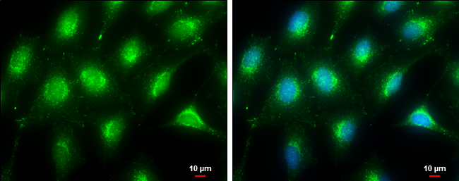
LAMP2 antibody detects LAMP2 protein at lysosome by immunofluorescent analysis.Sample: HeLa cells were fixed in ice-cold MeOH for 5 min.Green: LAMP2 stained by LAMP2 antibody (GTX103214) diluted at 1:500.Blue: Hoechst 33342 staining.Scale bar= 10μm.
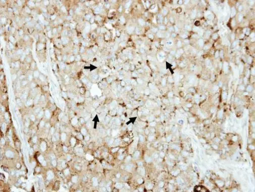
Immunohistochemical analysis of paraffin-embedded BT474 xenograft, using LAMP2(GTX103214) antibody at 1:500 dilution.
Antigen Retrieval: Trilogy™ (EDTA based, pH 8.0) buffer, 15min
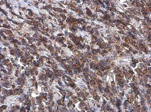
LAMP2 antibody detects LAMP2 protein at cytoplasm by immunohistochemical analysis.Sample: Paraffin-embedded human breast carcinoma.LAMP2 stained by LAMP2 antibody (GTX103214) diluted at 1:1000.Antigen Retrieval: Tris-EDTA buffer, pH 9.0, 15 min
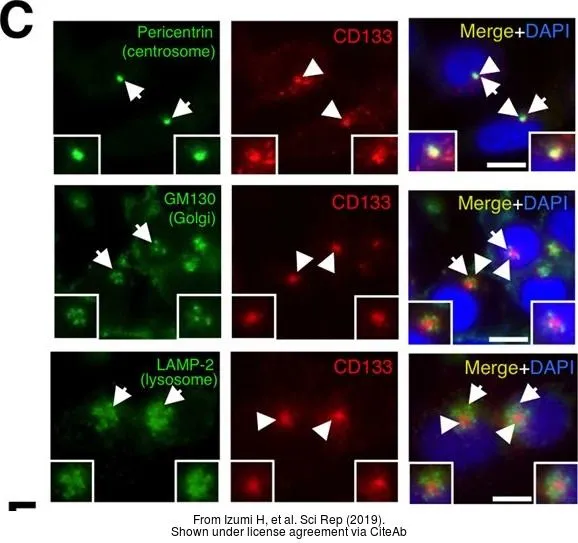
The data was published in the journal Sci Rep in 2019.PMID: 30783186
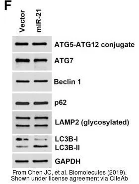
The data was published in the journal Biomolecules in 2019.PMID: 31505885
-
HostRabbit
-
ClonalityPolyclonal
-
IsotypeIgG
-
ApplicationsWB ICC/IF IHC-P
-
ReactivityHuman