LC3A antibody
Cat. No. GTX132889
Cat. No. GTX132889

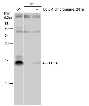
GTX132889 WB Image
RD and untreated (–) or treated (+) HeLa whole cell extracts (30 μg) were separated by 15% SDS-PAGE, and the membrane was blotted with LC3A antibody (GTX132889) diluted at 1:1000.
1 / 2
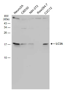
GTX132889 WB Image
Various whole cell extracts (30 μg) were separated by 15% SDS-PAGE, and the membrane was blotted with LC3A antibody (GTX132889) diluted at 1:1000. The HRP-conjugated anti-rabbit IgG antibody (GTX213110-01) was used to detect the primary antibody.
2 / 2
-
HostRabbit
-
ClonalityPolyclonal
-
IsotypeIgG
-
ApplicationsWB
-
ReactivityHuman, Mouse