LDB2 antibody
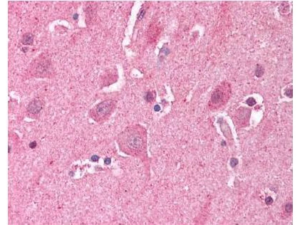
IHC-P analysis of human brain cortex tissue using GTX23627 LDB2 antibody.
Dilution : 5 μg/mL
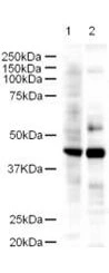
WB analysis of various samples using GTX23627 LDB2 antibody.
Lane 1 : Human kidney tissue lysate
Lane 2 : Mouse spleen tissue lysate
Loading : 18 μg
Dilution : 1:500
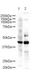
Western blot using GeneTex's Affinity Purified anti-LDB2 antibody shows detection of a 43-kDa band corresponding to LDB2 in a lysates prepared from human kidney (lane 1) and mouse spleen (lane 2) tissues. Approximately 18 μg of lysate was run on a SDS-PAGE and transferred onto nitrocellulose followed by reaction with a 1:500 dilution of anti-LDB2 antibody. Detection occurred using a 1:5,000 dilution of HRP-labeled Goat anti-Rabbit IgG for 1 hour at room temperature. A chemiluminescence system was used for signal detection (Roche) using a 1 min exposure time.
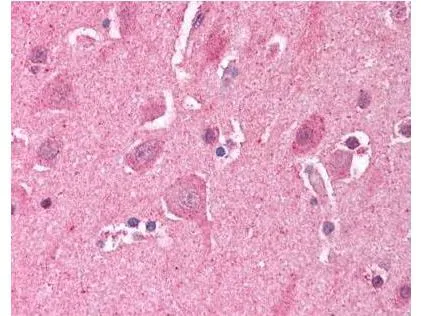
GeneTex's Affinity Purified anti-LDB2 (Clim1) antibody was used at a 5 μg/ml to detect LDB2 in human brain cortex tissue. The image shows the localization of antibody as the precipitated red signal, with a hematoxylin purple nuclear counter stain. Tissue was formalin-fixed and paraffin embedded.
-
HostRabbit
-
ClonalityPolyclonal
-
IsotypeIgG
-
ApplicationsWB IHC-P ELISA
-
ReactivityHuman, Mouse