LDHA antibody


Various whole cell extracts (30 μg) were separated by 12% SDS-PAGE, and the membrane was blotted with LDHA antibody (GTX101416) diluted at 1:1000. The HRP-conjugated anti-rabbit IgG antibody (GTX213110-01) was used to detect the primary antibody.

Various whole cell extracts (30 μg) were separated by 12% SDS-PAGE, and the membrane was blotted with LDHA antibody (GTX101416) diluted at 1:1000. The HRP-conjugated anti-rabbit IgG antibody (GTX213110-01) was used to detect the primary antibody.
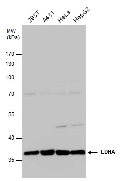
LDHA antibody detects LDHA protein by western blot analysis. Various whole cell extracts (30 μg) were separated by 10% SDS-PAGE, and the membrane was blotted with LDHA antibody (GTX101416) diluted at 1:1000. The HRP-conjugated anti-rabbit IgG antibody (GTX213110-01) was used to detect the primary antibody.
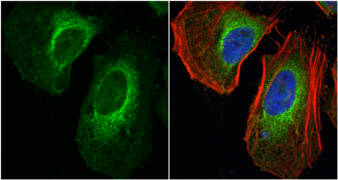
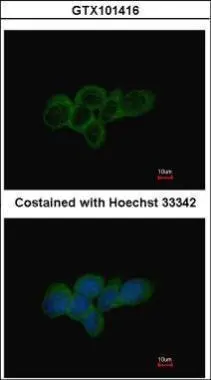
LDHA antibody detects LDHA protein at cytoplasm by immunofluorescent analysis.
Sample: HeLa cells were fixed in 4% paraformaldehyde at RT for 15 min.
Green: LDHA protein stained by LDHA antibody (GTX101416) diluted at 1:1000.
Red: phalloidin, a cytoskeleton marker, stained by phalloidin (invitrogen, A12380) diluted at 1:200.
Blue: Hoechst 33342 staining.
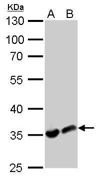
LDHA antibody detects LDHA protein by western blot analysis.
A. 30 μg PC-12 whole cell lysate/extract
B. 30 μg Rat2 whole cell lysate/extract
10% SDS-PAGE
LDHA antibody (GTX101416) dilution: 1:1000
The HRP-conjugated anti-rabbit IgG antibody (GTX213110-01) was used to detect the primary antibody.
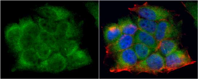
LDHA antibody detects LDHA protein at cytoplasm by immunofluorescent analysis.
Sample: MCF7 cells were fixed in 4% paraformaldehyde at RT for 15 min.
Green: LDHA protein stained by LDHA antibody (GTX101416) diluted at 1:1000.
Red: phalloidin, a cytoskeleton marker, stained by phalloidin (invitrogen, A12380) diluted at 1:200.
Blue: Hoechst 33342 staining.
Immunofluorescence analysis of methanol-fixed A431, using LDHA(GTX101416) antibody at 1:500 dilution.
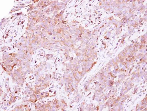
Immunohistochemical analysis of paraffin-embedded human lung papillory adenocarcinoma, using LDHA(GTX101416) antibody at 1:500 dilution.
Antigen Retrieval: Trilogy™ (EDTA based, pH 8.0) buffer, 15min
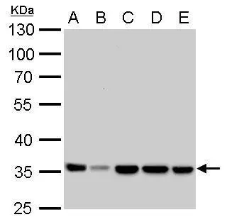
LDHA antibody detects LDHA protein by western blot analysis.
A. 30 μg Neuro2A whole cell lysate/extract
B. 30 μg C8D30 whole cell lysate/extract
C. 30 μg NIH-3T3 whole cell lysate/extract
D. 30 μg Raw264.7 whole cell lysate/extract
E. 30 μg C2C12 whole cell lysate/extract
10% SDS-PAGE
LDHA antibody (GTX101416) dilution: 1:1000
The HRP-conjugated anti-rabbit IgG antibody (GTX213110-01) was used to detect the primary antibody.

LDHA antibody detects LDHA protein at cytoplasm by immunohistochemical analysis.Sample: Paraffin-embedded mouse liver.LDHA stained by LDHA antibody (GTX101416) diluted at 1:500.Antigen Retrieval: Citrate buffer, pH 6.0, 15 min
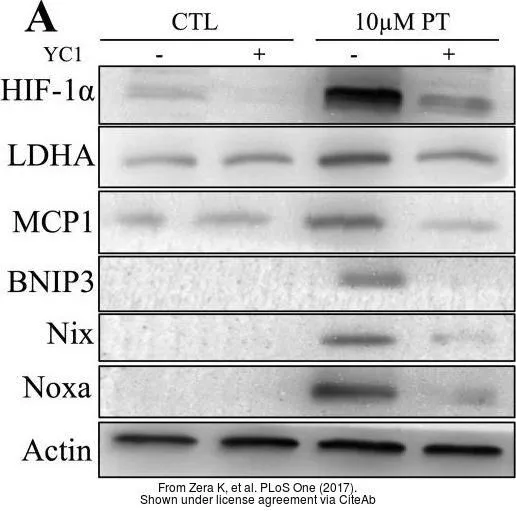
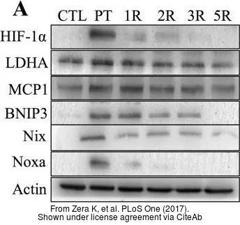
The data was published in the journal PLoS One in 2017. PMID: 29045486
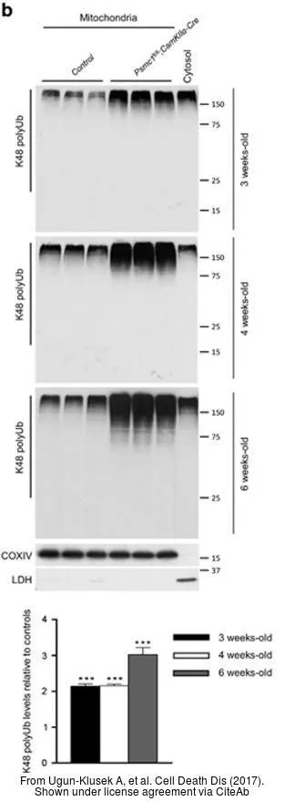
The data was published in the journal Cell Death Dis in 2017. PMID: 28055010
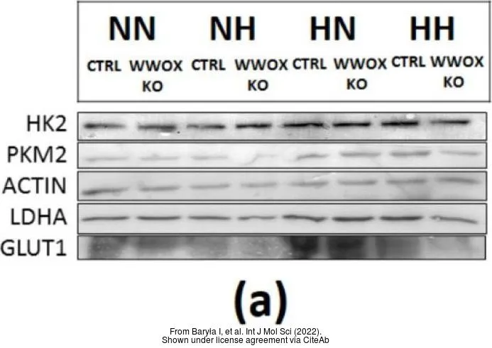
The data was published in the 2022 in Int J Mol Sci. PMID: 35328751
-
HostRabbit
-
ClonalityPolyclonal
-
IsotypeIgG
-
ApplicationsWB ICC/IF IHC-P
-
ReactivityHuman, Mouse, Rat