LEF1 antibody, Internal
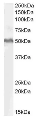
WB analysis of MOLT4 whole cell lysate using GTX89284 LEF1 antibody, Internal.
Dilution : 1 μg/ml
Loading : 35μg protein in RIPA buffer
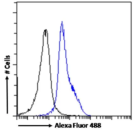
FACS analysis of PFA-fixed Jurkat cells using GTX89284 LEF1 antibody, Internal.
Blue : Primary antibody
Black : Unimmunized goat IgG
Permeabilization : 0.5% Triton
Dilution : 10μg/ml
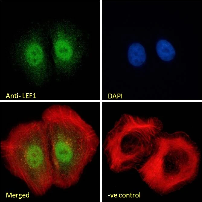
ICC/IF analysis of PFA-fixed U2OS cells using GTX89284 LEF1 antibody, Internal. Actin filaments were stained with phalloidin (red) and the nuclear stain is DAPI (blue).
Negative control (-ve) : Unimmunized goat IgG
Permeabilization : 0.15% Triton
Dilution : 10μg/ml
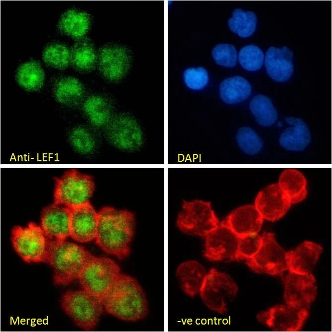
ICC/IF analysis of PFA-fixed Jurkat cells using GTX89284 LEF1 antibody, Internal. Actin filaments were stained with phalloidin (red) and the nuclear stain is DAPI (blue).
Negative control (-ve) : Unimmunized goat IgG
Permeabilization : 0.15% Triton
Dilution : 10μg/ml
-
HostGoat
-
ClonalityPolyclonal
-
IsotypeIgG
-
ApplicationsWB ICC/IF FCM
-
ReactivityHuman