LMO1 antibody
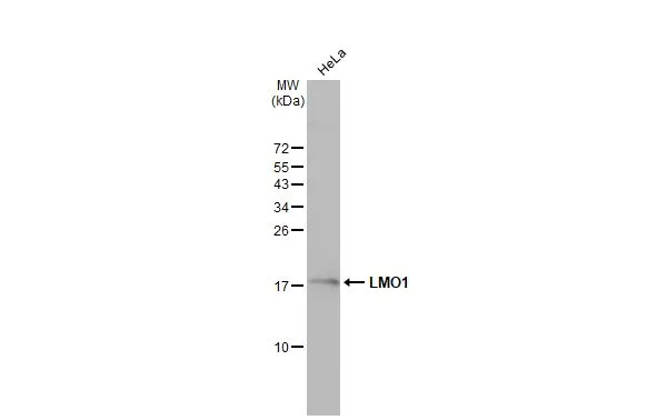
Whole cell extract (30 μg) was separated by 15% SDS-PAGE, and the membrane was blotted with LMO1 antibody (GTX101726) diluted at 1:1000. The HRP-conjugated anti-rabbit IgG antibody (GTX213110-01) was used to detect the primary antibody.
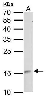
LMO1 antibody detects LMO1 protein by Western blot analysis.
A. 50 μg rat brain lysate/extract
12 % SDS-PAGE
LMO1 antibody (GTX101726) dilution: 1:500
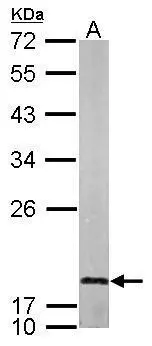
Sample (50 ug of whole cell lysate)
A: mouse brain
12% SDS PAGE
GTX101726 diluted at 1:1000
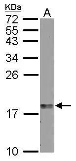
Sample (30 μg of whole cell lysate)
A: zebrafish brain
15% SDS PAGE
GTX101726 diluted at 1:1000
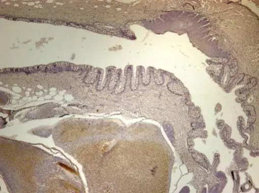
Immunohistochemical analysis of paraffin-embedded zebrafish tissue, using LMO1 antibody (GTX101726) at 1:300 dilution.
-
HostRabbit
-
ClonalityPolyclonal
-
IsotypeIgG
-
ApplicationsWB IHC-P
-
ReactivityHuman, Mouse, Rat, Zebrafish