Lamin A + C antibody

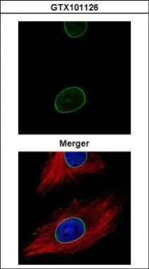
Confocal immunofluorescence analysis (Olympus FV10i) of methanol-fixed HeLa, using Lamin A + C (GTX101126) antibody (Green) at 1:500 dilution. Alpha-tubulin filaments were labeled with GTX11304 (Red) at 1:500.
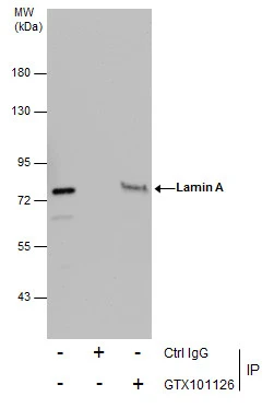
Immunoprecipitation of Lamin A + C protein from HeLa whole cell extracts using 5 μg of Lamin A + C antibody (GTX101126).
Western blot analysis was performed using Lamin A + C antibody (GTX101126).
EasyBlot anti-Rabbit IgG (GTX221666-01) was used as a secondary reagent.
Lamin A + C antibody detects Lamin A + C protein at nuclear envelope on mouse prostate by immunohistochemical analysis.
Sample: Paraffin-embedded mouse prostate.
Lamin A + C antibody (GTX101126) dilution: 1:500.
Antigen Retrieval: Trilogy™ (EDTA based, pH 8.0) buffer, 15min
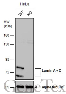
Wild-type (WT) and Lamin A + C knockout (KO) HeLa cell extracts (30 μg) were separated by 7.5% SDS-PAGE, and the membrane was blotted with Lamin A + C antibody (GTX101126) diluted at 1:5000. The HRP-conjugated anti-rabbit IgG antibody (GTX213110-01) was used to detect the primary antibody, and the signal was developed with Trident ECL plus-Enhanced.
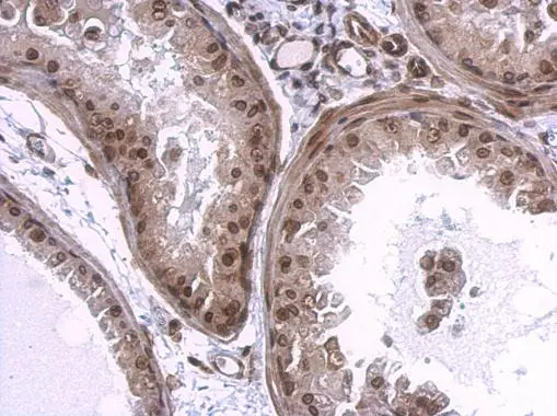
Lamin A + C antibody detects Lamin A + C protein at nuclear envelope on human breast carcinoma by immunohistochemical analysis.
Sample: Paraffin-embedded human breast carcinoma.
Lamin A + C antibody (GTX101126) dilution: 1:500.
Antigen Retrieval: Trilogy™ (EDTA based, pH 8.0) buffer, 15min
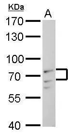
Lamin A + C antibody detects Lamin A + C protein by Western blot analysis.
A. 30 μg C2C12 whole cell lysate/extract
7.5 % SDS-PAGE
Lamin A + C antibody (GTX101126) dilution: 1:1000
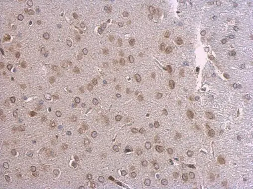
Lamin A + C antibody detects Lamin A + C protein at nuclear envelope on mouse fore brain by immunohistochemical analysis.
Sample: Paraffin-embedded mouse fore brain.
Lamin A + C antibody (GTX101126) dilution: 1:500.
Antigen Retrieval: Trilogy™ (EDTA based, pH 8.0) buffer, 15min
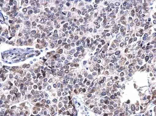
Lamin A + C antibody detects Lamin A + C protein at membrane on U87 xenograft by immunohistochemical analysis.
Sample: Paraffin-embedded U87 xenograft.
Lamin A + C antibody (GTX101126) dilution: 1:500.
Antigen Retrieval: Trilogy™ (EDTA based, pH 8.0) buffer, 15min
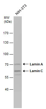
Whole cell extract (30 μg) was separated by 7.5% SDS-PAGE, and the membrane was blotted with Lamin A + C antibody (GTX101126) diluted at 1:1000.
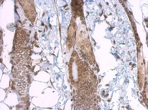
Lamin A + C antibody detects Lamin A + C protein at nuclear envelope on mouse skin by immunohistochemical analysis.
Sample: Paraffin-embedded mouse skin.
Lamin A + C antibody (GTX101126) dilution: 1:500.
Antigen Retrieval: Trilogy™ (EDTA based, pH 8.0) buffer, 15min
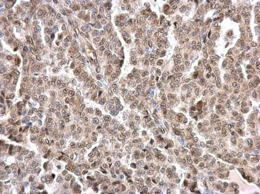
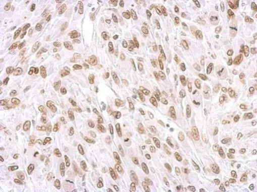
Lamin A + C antibody detects Lamin A + C protein at nuclear envelope on human endometrial carcinoma by immunohistochemical analysis.
Sample: Paraffin-embedded human endometrial carcinoma.
Lamin A + C antibody (GTX101126) dilution: 1:500.
Antigen Retrieval: Trilogy™ (EDTA based, pH 8.0) buffer, 15min
-
HostRabbit
-
ClonalityPolyclonal
-
IsotypeIgG
-
ApplicationsWB ICC/IF IHC-P IP
-
ReactivityHuman, Mouse