Lamin A + C antibody
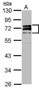
Sample (20 ug )
A: HeLa Nucleus
10% SDS PAGE
GTX111677 diluted at 1:3000
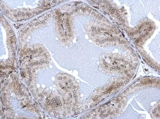
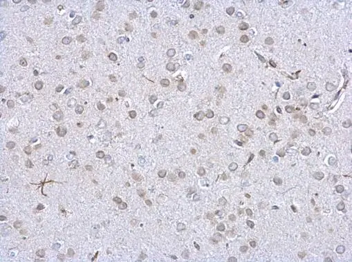
Lamin A + C antibody detects Lamin A + C protein at nuclear envelope on mouse prostate by immunohistochemical analysis.
Sample: Paraffin-embedded mouse prostate.
Lamin A + C antibody (GTX111677) dilution: 1:500.
Antigen Retrieval: Trilogy™ (EDTA based, pH 8.0) buffer, 15min
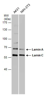
Various whole cell extracts (30 μg) were separated by 7.5% SDS-PAGE, and the membrane was blotted with Lamin A + C antibody (GTX111677) diluted at 1:1000.
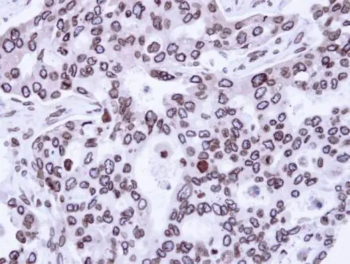
Immunohistochemical analysis of paraffin-embedded H441 Xenograft, using Lamin A + C(GTX111677) antibody at 1:100 dilution.
Antigen Retrieval: Trilogy™ (EDTA based, pH 8.0) buffer, 15min
Lamin A + C antibody detects Lamin A + C protein at nuclear envelope on mouse prostate by immunohistochemical analysis.
Sample: Paraffin-embedded mouse prostate.
Lamin A + C antibody (GTX111677) dilution: 1:500.
Antigen Retrieval: Trilogy™ (EDTA based, pH 8.0) buffer, 15min
-
HostRabbit
-
ClonalityPolyclonal
-
IsotypeIgG
-
ApplicationsWB IHC-P
-
ReactivityHuman, Mouse