MAD2L2 antibody

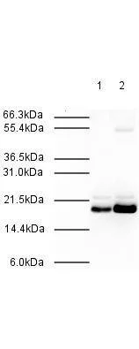
WB analysis of HeLa whole cell lysate using GTX23630 MAD2L2 antibody.
Lane 1 : whole cell lysate
Lane 2 : Nuclear extract
Loading : 20 μg
Dilution : 1:500
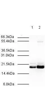
Affinity Purified Rabbit anti-MAD2L2 was used at a 1:500 dilution to detect human MAD2L2 by western blot. Both HeLa whole cell lysate (lane 1) and nuclear lysate (lane 2) were probed using this antibody. This antibody clearly detects a ~20 kDa band corresponding to human MAD2L2 (predicted molecular weight is 24 kDa). Approximately 20 μg of each lysate was loaded on a 10% SDS-PAGE. Primary antibody was reacted with the membrane at room temperature for 1 h. After subsequent washing, a 1:2,000 dilution of HRP conjugated Gt-a-Rabbit IgG was used for visualization. Exposure time was 30 sec.
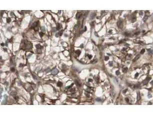
GeneTex Affinity Purified anti-MAD2L2 antibody (GTX23630) shows strong nuclear and cytoplasmic staining of tumor cells in cancerous human kidney tissue. Tissue was formalin-fixed and paraffin embedded. Brown color indicates presence of protein, blue color shows cell nuclei.
-
HostRabbit
-
ClonalityPolyclonal
-
IsotypeIgG
-
ApplicationsWB IHC-P ELISA
-
ReactivityHuman