MAFF antibody


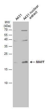
A431 whole cell and nuclear extracts (30 μg) were separated by 15% SDS-PAGE, and the membrane was blotted with MAFF antibody (GTX120264) diluted at 1:500. The HRP-conjugated anti-rabbit IgG antibody (GTX213110-01) was used to detect the primary antibody.
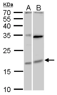
MAFF antibody detects MAFF protein by Western blot analysis.
A. 30 μg Huh7 whole cell lysate/extract
B. 30 μg HepG2 whole cell lysate/extract
12 % SDS-PAGE
MAFF antibody (GTX120264) dilution: 1:2000
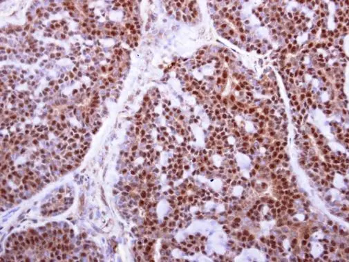
MAFF antibody detects MAFF protein at cytoplasm and nucleus on human breast carcinoma by immunohistochemical analysis.
Sample: Paraffin-embedded human breast carcinoma.
MAFF antibody (GTX120264) diluted at 1:250.
Antigen Retrieval: Trilogy™ (EDTA based, pH 8.0) buffer, 15min
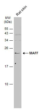
Rat tissue extract (50 μg) was separated by 15% SDS-PAGE, and the membrane was blotted with MAFF antibody (GTX120264) diluted at 1:500. The HRP-conjugated anti-rabbit IgG antibody (GTX213110-01) was used to detect the primary antibody.
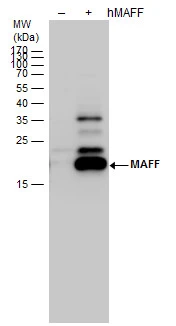
MAFF antibody detects MAFF protein by western blot analysis. Non-transfected (-) and MAFF -transfected (+, ) 293T whole cell extracts (30 μg) were separated by 12% SDS-PAGE, and the membrane was blotted with MAFF antibody (GTX120264) diluted by 1:20000.
-
HostRabbit
-
ClonalityPolyclonal
-
IsotypeIgG
-
ApplicationsWB IHC-P
-
ReactivityHuman, Rat