MDM2 (phospho Ser185) antibody

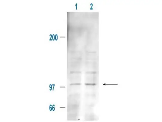
WB analysis of 293T whole cell lysate using GTX21094 MDM2 (phospho Ser185) antibody.
Lane 1 : Untreated whole cell lysate
Lane 2 : 100 ng/mL IGF-1 treated whole cell lysate
Dilution : 20 μg
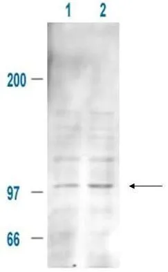
Affinity Purified Anti-MDM2 pS185 (Rabbit) is shown to detect a 102 kDa band (arrow) corresponding to phosphorylated mouse MDM2 present in a 293T whole cell lysate. Cells were serum-starved for 24 hours prior to harvest. Approxi-mately 20 ug of lysate was loaded per lane for SDS-PAGE. Untreated cells are shown in lane 1, whereas cells in lane 2 were treated with IGF-1 (100 ng/ml) for 20 min prior to harvest. Follow reaction of antibody with a 1:2000 dilution of HRP Goat-a-Rabbit IgG for visualization.
-
HostRabbit
-
ClonalityPolyclonal
-
IsotypeIgG
-
ApplicationsWB IP ELISA
-
ReactivityHuman, Mouse