MEF2C antibody


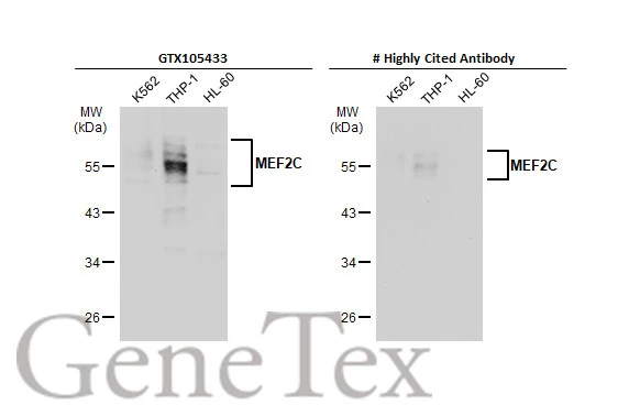
Various whole cell extracts (30 μg) were separated by 10% SDS-PAGE, and the membranes were blotted with MEF2C antibody (GTX105433) diluted at 1:1000 and competitor's antibody diluted at 1:1000. The HRP-conjugated anti-rabbit IgG antibody (GTX213110-01) was used to detect the primary antibody.
*The competitor is not affiliated with GeneTex and does not endorse this product.
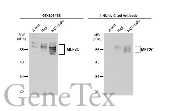
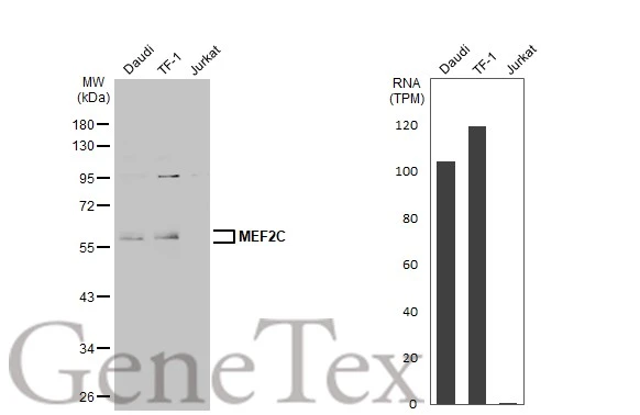
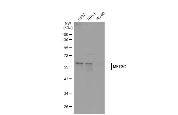
Various whole cell extracts (30 μg) were separated by 10% SDS-PAGE, and the membrane was blotted with MEF2C antibody (GTX105433) diluted at 1:1000. The HRP-conjugated anti-rabbit IgG antibody (GTX213110-01) was used to detect the primary antibody. Corresponding RNA expression data for the same cell lines are based on Human Protein Atlas program.
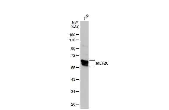
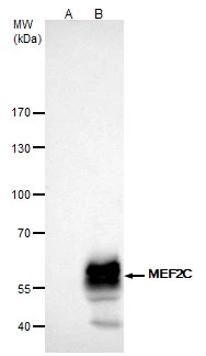
Whole cell extract (30 μg) was separated by 10% SDS-PAGE, and the membrane was blotted with MEF2C antibody (GTX105433) diluted at 1:1000. The HRP-conjugated anti-rabbit IgG antibody (GTX213110-01) was used to detect the primary antibody.
MEF2C antibody detects MEF2C protein at nucleus on Saos2 xenograft by immunohistochemical analysis.
Sample: Paraffin-embedded Saos2 xenograft.
MEF2C antibody (GTX105433) dilution: 1:500.
Antigen Retrieval: Trilogy™ (EDTA based, pH 8.0) buffer, 15min
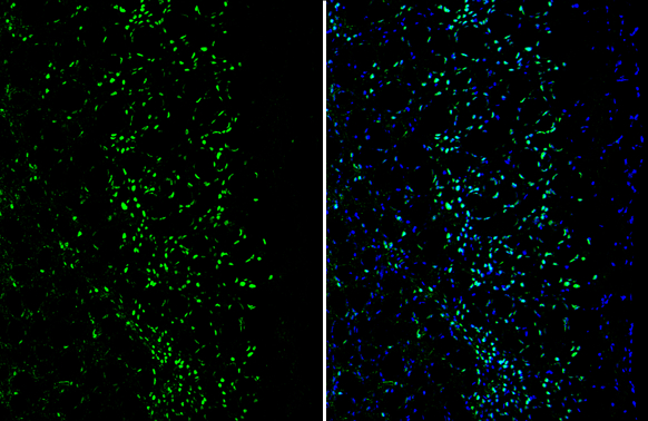
MEF2C antibody detects MEF2C protein by immunohistochemical analysis.Sample: Frozen-sectioned mouse cerebral cortex.Green: MEF2C stained by MEF2C antibody (GTX105433) diluted at 1:250.Blue: Fluoroshield with DAPI (GTX30920).
MEF2C antibody immunoprecipitates MEF2C protein in IP experiments.
IP samples: K562 whole cell extract
A. Control with 4 μg of preimmune Rabbit IgG
B. Immunoprecipitation of MEF2C protein by 4 μg MEF2C antibody (GTX105433)
5 % SDS-PAGE
The immunoprecipitated MEF2C protein was detected by MEF2C antibody (GTX105433) diluted at 1:1000.
[EasyBlot anti-rabbit IgG (GTX221666-01) was used as a secondary reagent]

MEF2C antibody detects MEF2C protein at cytoplasm and nucleus by immunofluorescent analysis.
Sample: HeLa cells were fixed in 4% paraformaldehyde at RT for 15 min.
Green: MEF2C protein stained by MEF2C antibody (GTX105433) diluted at 1:1000.
Red: phalloidin, a cytoskeleton marker, diluted at 1:200.
Scale bar = 10 μm.
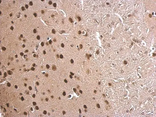
MEF2C antibody detects MEF2C protein at nucleus on mouse fore brain by immunohistochemical analysis.
Sample: Paraffin-embedded mouse fore brain.
MEF2C antibody (GTX105433) dilution: 1:500.
Antigen Retrieval: Trilogy™ (EDTA based, pH 8.0) buffer, 15min
Various whole cell extracts (30 μg) were separated by 10% SDS-PAGE, and the membrane was blotted with MEF2C antibody (GTX105433) diluted at 1:1000. The HRP-conjugated anti-rabbit IgG antibody (GTX213110-01) was used to detect the primary antibody.
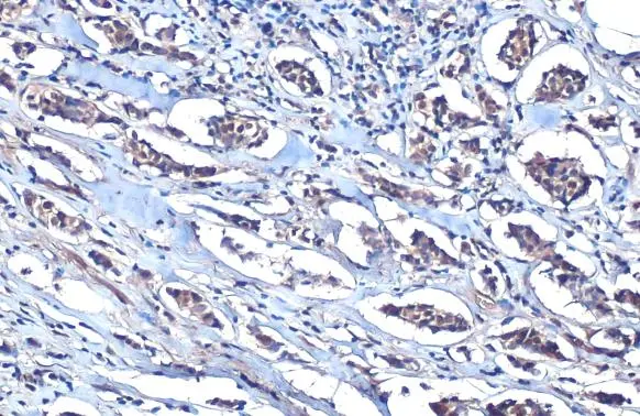
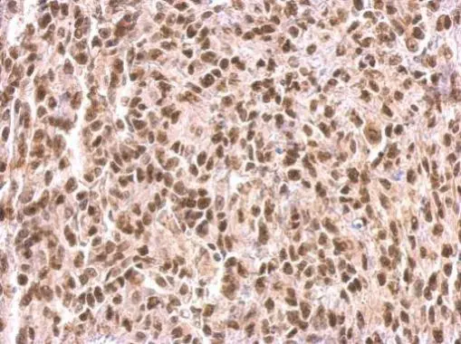
MEF2C antibody detects MEF2C protein at cytoplasm and nucleus by immunohistochemical analysis.Sample: Paraffin-embedded human breast carcinoma.MEF2C stained by MEF2C antibody (GTX105433) diluted at 1:250.Antigen Retrieval: Citrate buffer, pH 6.0, 15 min
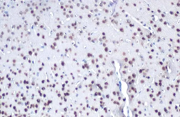
MEF2C antibody detects MEF2C protein at nucleus by immunohistochemical analysis.Sample: Paraffin-embedded mouse brain.MEF2C stained by MEF2C antibody (GTX105433) diluted at 1:250.Antigen Retrieval: Citrate buffer, pH 6.0, 15 min
-
HostRabbit
-
ClonalityPolyclonal
-
IsotypeIgG
-
ApplicationsWB ICC/IF IHC-P IHC-Fr IP
-
ReactivityHuman, Mouse