MIP3 alpha antibody
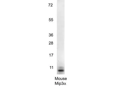
WB analysis of recombinant mouse MIP3 alpha protein using GTX48690 MIP3 alpha antibody.
Loading : 0.25 μg
Dilution : 1:1000
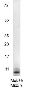
Western blot using GeneTex's anti-Mouse MIP3a antibody shows detection of a band ~11 kDa in size corresponding to recombinant mouse MIP3a. Recombinant mouse MIP-3a was loaded on to an SDS-PAGE gel at 0.25 μg and after separation was transferred to nitrocellulose. The membrane was blocked with 1% BSA in TBST for 30 min at RT, followed by incubation with primary antibody diluted 1:1,000 in 1% BSA in TBST overnight at 4ºC. After washes, the blot was reacted with secondary antibody HRP Goat anti-Rabbit IgG antibody diluted 1:40,000 in blocking buffer) for 30 min at RT followed by reation with FemtoMax Chemiluminescent substrate. Data was collected using Bio-Rad VersaDocR 4000 MP imaging system.
-
HostRabbit
-
ClonalityPolyclonal
-
IsotypeIgG
-
ApplicationsWB ELISA
-
ReactivityMouse, Rat