APPLICATION
Application Note
*Optimal dilutions/concentrations should be determined by the researcher.
Application |
Recommended Dilution |
1:500-1:3000 |
1:100-1:1000 |
Not tested in other applications.
Calculated MW
Positive Control
U87-MG , SK-N-SH , IMR32 , SK-N-AS
Predict Reactivity
Bovine(>80% identity)
PROPERTIES
Form
Liquid
Buffer
0.1M Tris, 0.1M Glycine, 20% Glycerol
Preservative
0.01% Thimerosal
Storage
Store as concentrated solution. Centrifuge briefly prior to opening vial. For short-term storage (1-2 weeks), store at 4ºC. For long-term storage, aliquot and store at -20ºC or below. Avoid multiple freeze-thaw cycles.
Concentration
0.69 mg/ml (Please refer to the vial label for the specific concentration.)
Antigen Species
Human
Immunogen
Recombinant protein encompassing a sequence within the center region of human MRPL42. The exact sequence is proprietary.
Purification
Purified by antigen-affinity chromatography.
Conjugation
Unconjugated
RRID
AB_2885475
Note
For laboratory research use only. Not for any clinical, therapeutic, or diagnostic use in humans or animals. Not for animal or human consumption.
Purchasers shall not, and agree not to enable third parties to, analyze, copy, reverse engineer or otherwise attempt to determine the structure or sequence of the product.
TARGET
Synonyms
mitochondrial ribosomal protein L42 , HSPC204 , L31MT , L42MT , MRP-L31 , MRP-L42 , MRP-S32 , MRPL31 , MRPS32 , PTD007 , RPML31 , S32MT
Cellular Localization
Mitochondrion
Background
Mammalian mitochondrial ribosomal proteins are encoded by nuclear genes and help in protein synthesis within the mitochondrion. Mitochondrial ribosomes (mitoribosomes) consist of a small 28S subunit and a large 39S subunit. They have an estimated 75% protein to rRNA composition compared to prokaryotic ribosomes, where this ratio is reversed. Another difference between mammalian mitoribosomes and prokaryotic ribosomes is that the latter contain a 5S rRNA. Among different species, the proteins comprising the mitoribosome differ greatly in sequence, and sometimes in biochemical properties, which prevents easy recognition by sequence homology. This gene encodes a protein identified as belonging to both the 28S and the 39S subunits. Further experiments will be needed to identify the specific subunit localization. Sequence analysis identified three transcript variants that encode two different isoforms. Pseudogenes corresponding to this gene are found on chromosomes 4q, 6p, 6q, 7p, and 15q. [provided by RefSeq]
Database
Research Area
DATA IMAGES
|
GTX120260 WB Image
MRPL42 antibody detects MRPL42 protein by Western blot analysis. A. 30 μg U87-MG whole cell lysate/extract B. 30 μg SK-N-SH whole cell lysate/extract C. 30 μg IMR32 whole cell lysate/extract D. 30 μg SK-N-AS whole cell lysate/extract 12 % SDS-PAGE MRPL42 antibody (GTX120260) dilution: 1:1000
|
|
GTX120260 ICC/IF Image
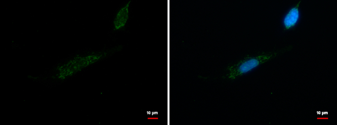
MRPL42 antibody detects MRPL42 protein at mitochondrion by immunofluorescent analysis. Sample: SK-N-SH cells were fixed in 100% MeOH for 5 min. Green: MRPL42 protein stained by MRPL42 antibody (GTX120260) diluted at 1:200. Blue: Hoechst 33342 staining.
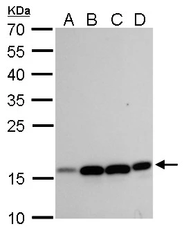
|
REFERENCE
There are currently no references for MRPL42 antibody (GTX120260). Be the first to share your publications with this product.
REVIEW
There are currently no reviews for MRPL42 antibody (GTX120260). Be the first to share your experience with this product.