Munc18-1 antibody

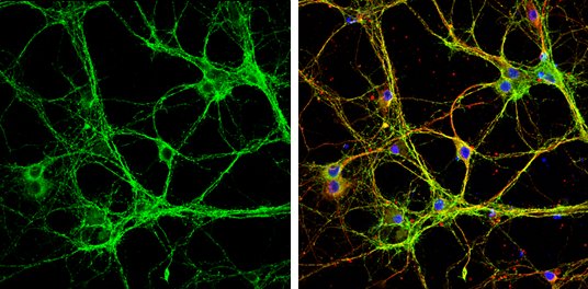
Munc18-1 antibody detects Munc18-1 protein by immunofluorescent analysis.
Sample: DIV9 rat E18 primary cortical neurons were fixed in 4% paraformaldehyde at RT for 15 min.
Green: Munc18-1 protein stained by Munc18-1 antibody (GTX114809) diluted at 1:500.
Red: beta Tubulin 3/ Tuj1, stained by beta Tubulin 3/ Tuj1 antibody [GT886] (GTX631830) diluted at 1:500.
Blue: Fluoroshield with DAPI (GTX30920).
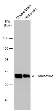
Various tissue extracts (50 μg) were separated by 7.5% SDS-PAGE, and the membrane was blotted with Munc18-1 antibody (GTX114809) diluted at 1:10000. The HRP-conjugated anti-rabbit IgG antibody (GTX213110-01) was used to detect the primary antibody.
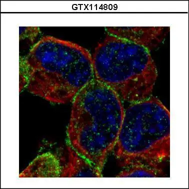
Confocal immunofluorescence analysis (Olympus FV10i) of methanol-fixed A431, using STXBP1(GTX114809) antibody (Green) at 1:500 dilution. Alpha-tubulin filaments were labeled with GTX11304 (Red) at 1:2000.
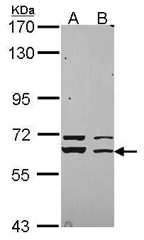
Sample (30 ug of whole cell lysate)
A: 293T
B: A431 (GTX27909)
7.5% SDS PAGE
GTX114809 diluted at 1:3000
-
HostRabbit
-
ClonalityPolyclonal
-
IsotypeIgG
-
ApplicationsWB ICC/IF IP
-
ReactivityHuman, Mouse, Rat