NDP52 antibody


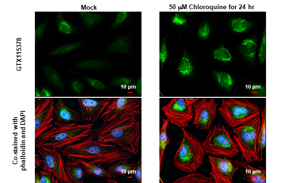
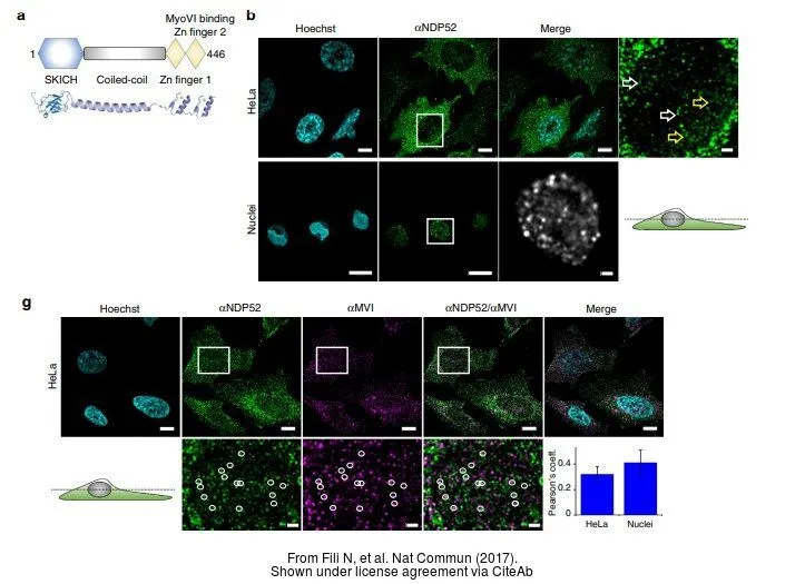
NDP52 antibody detects NDP52 protein at autophagosome by immunofluorescent analysis.Sample: Mock and treated HeLa cells were fixed in 4% paraformaldehyde at RT for 15 min.Green: NDP52 stained by NDP52 antibody (GTX115378) diluted at 1:1000.Red: phalloidin, a cytoskeleton marker, diluted at 1:200.Blue: Fluoroshield with DAPI (GTX30920).Scale bar= 10 μm.

Untreated (–) and treated (+) HepG2 whole cell extracts (30 μg) were separated by 10% SDS-PAGE, and the membrane was blotted with NDP52 antibody (GTX115378) diluted at 1:2000. The HRP-conjugated anti-rabbit IgG antibody (GTX213110-01) was used to detect the primary antibody.
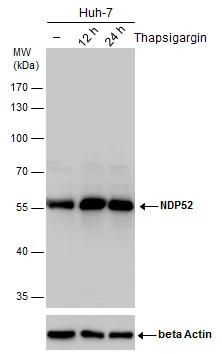
NDP52 antibody detects NDP52 protein by western blot analysis. Un-treated (-) and treated (+, Thapsigargin treatment for 12hrs and 24hrs) Huh-7 whole cell extracts (30 μg) were separated by 10% SDS-PAGE, and the membrane was blotted with NDP52 antibody (GTX115378) diluted by 1:2000.
The ACTB was used as internal control (GTX110564, 1:50000) shown at the bottom panel.
The HRP-conjugated anti-rabbit IgG antibody (GTX213110-01) was used to detect the primary antibody.
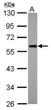
Sample (30 μg of whole cell lysate)
A: Raji
10% SDS PAGE
GTX115378 diluted at 1:1000
The HRP-conjugated anti-rabbit IgG antibody (GTX213110-01) was used to detect the primary antibody.
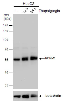
NDP52 antibody detects NDP52 protein by western blot analysis. Un-treated (-) and treated (+, Thapsigargin treatment for 12hrs and 24hrs) HepG2 whole cell extracts (30 μg) were separated by 10% SDS-PAGE, and the membrane was blotted with NDP52 antibody (GTX115378) diluted by 1:2000.
The ACTB was used as internal control (GTX110564, 1:50000) shown at the bottom panel.
The HRP-conjugated anti-rabbit IgG antibody (GTX213110-01) was used to detect the primary antibody.
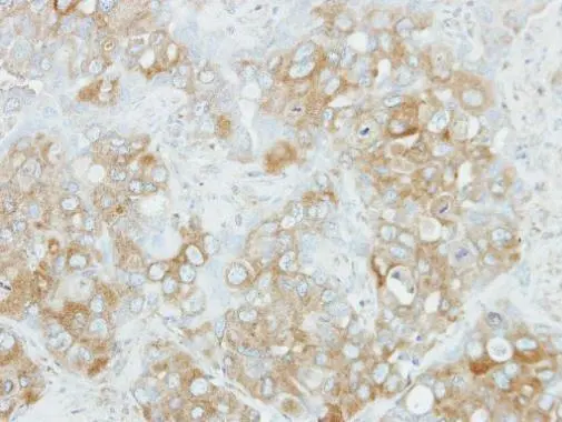
Immunohistochemical analysis of paraffin-embedded H441 xenograft, using NDP52(GTX115378) antibody at 1:100 dilution.
Antigen Retrieval: Trilogy™ (EDTA based, pH 8.0) buffer, 15min
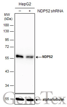
Non-transfected (–) and transfected (+) HepG2 whole cell extracts (30 μg) were separated by 10% SDS-PAGE, and the membrane was blotted with NDP52 antibody (GTX115378) diluted at 1:4000. The HRP-conjugated anti-rabbit IgG antibody (GTX213110-01) was used to detect the primary antibody.
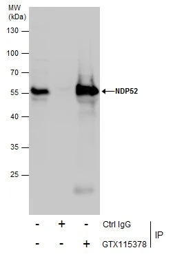
Immunoprecipitation of NDP52 protein from Jurkat whole cell extracts using 5 μg of NDP52 antibody (GTX115378).
Western blot analysis was performed using NDP52 antibody (GTX115378).
EasyBlot anti-Rabbit IgG (GTX221666-01) was used as a secondary reagent.
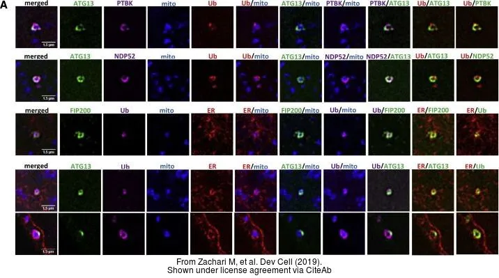
The data was published in the journal Dev Cell in 2019.PMID: 31353311
-
HostRabbit
-
ClonalityPolyclonal
-
IsotypeIgG
-
ApplicationsWB ICC/IF IHC-P IP
-
ReactivityHuman