NDUFA9 antibody

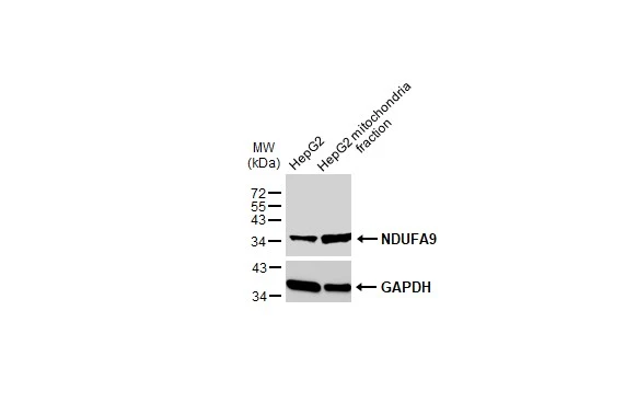
HepG2 and mitochondria extracts (30 μg) were separated by SDS-PAGE, and the membrane was blotted with NDUFA9 antibody (GTX132726) diluted at 1:1000. The HRP-conjugated anti-rabbit IgG antibody (GTX213110-01) was used to detect the primary antibody.
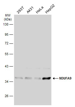
Various whole cell extracts (30 μg) were separated by 10% SDS-PAGE, and the membrane was blotted with NDUFA9 antibody (GTX132726) diluted at 1:1000. The HRP-conjugated anti-rabbit IgG antibody (GTX213110-01) was used to detect the primary antibody.
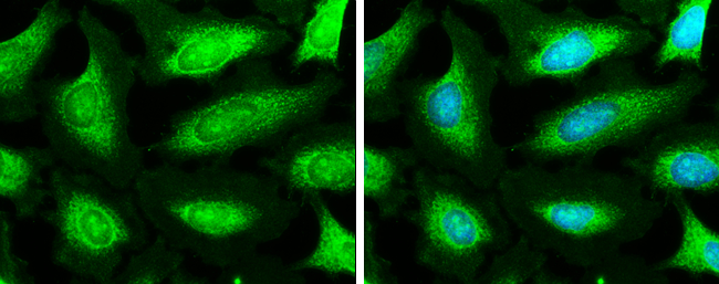
NDUFA9 antibody detects NDUFA9 protein at mitochondria by immunofluorescent analysis.Sample: HeLa cells were fixed in ice-cold MeOH for 5 min.Green: NDUFA9 stained by NDUFA9 antibody (GTX132726) diluted at 1:500.Blue: Hoechst 33342 staining.
-
HostRabbit
-
ClonalityPolyclonal
-
IsotypeIgG
-
ApplicationsWB ICC/IF
-
ReactivityHuman