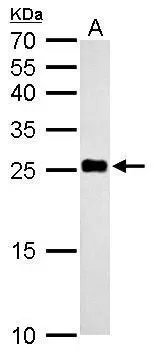
NDUFV2 antibody
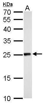
NDUFV2 antibody detects NDUFV2 protein by Western blot analysis.
A. 50 μg rat liver lysate/extract
12 % SDS-PAGE
NDUFV2 antibody (GTX102259) dilution: 1:500
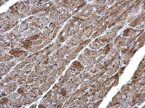
NDUFV2 antibody detects NDUFV2 protein at mitochondria on mouse heart by immunohistochemical analysis.
Sample: Paraffin-embedded mouse heart.
NDUFV2 antibody (GTX102259) diluted at 1:1000.
Antigen Retrieval: Trilogy™ (EDTA based, pH 8.0) buffer, 15min
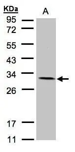
Sample(30 μg of whole cell lysate)
A:A431(GTX27909)
12% SDS PAGE
GTX102259 diluted at 1:1500
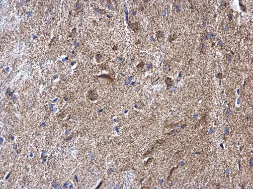
NDUFV2 antibody detects NDUFV2 protein at mitochondria on mouse heart by immunohistochemical analysis.
Sample: Paraffin-embedded mouse heart.
NDUFV2 antibody (GTX102259) diluted at 1:1000.
Antigen Retrieval: Trilogy™ (EDTA based, pH 8.0) buffer, 15min
NDUFV2 antibody detects NDUFV2 protein by Western blot analysis.
A. 50 μg mouse liver lysate/extract
12 % SDS-PAGE
NDUFV2 antibody (GTX102259) dilution: 1:500
-
HostRabbit
-
ClonalityPolyclonal
-
IsotypeIgG
-
ApplicationsWB IHC-P
-
ReactivityHuman, Mouse, Rat