NFAT1 antibody, Internal
Cat. No. GTX88316
Cat. No. GTX88316
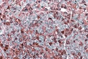
GTX88316 IHC-P Image
IHC-P analysis of human spleen using GTX88316 NFAT1 antibody, Internal.
Antigen retrieval : citrate buffer pH 6
Dilution : 3.8μg/ml
1 / 2
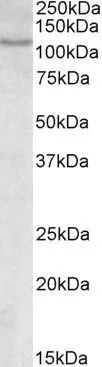
GTX88316 WB Image
WB analysis of Molt-4 cell lysate using GTX88316 NFAT1 antibody, Internal.
Dilution : 0.3μg/ml
Loading : 35μg protein in RIPA buffer
2 / 2
-
HostGoat
-
ClonalityPolyclonal
-
IsotypeIgG
-
ApplicationsWB IHC-P
-
ReactivityHuman