NFAT1 antibody

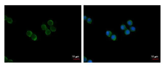
NFAT1 antibody detects NFAT1 protein at cytoplasm by immunofluorescent analysis.
Sample: RAW264.7 cells were fixed in 4% paraformaldehyde at RT for 15 min.
Green: NFAT1 protein stained by NFAT1 antibody (GTX127932) diluted at 1:500.
Blue: Hoechst 33342 staining.
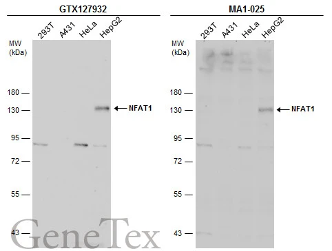
Various whole cell extracts (30 μg) were separated by 7.5% SDS-PAGE, and the membranes were blotted with NFAT1 antibody (GTX127932) diluted at 1:500 and competitor's antibody (MA1-025) diluted at 1:500. The HRP-conjugated anti-rabbit IgG antibody (GTX213110-01) was used to detect the primary antibody.
*The competitor is not affiliated with GeneTex and does not endorse this product.
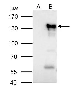
NFAT1 antibody immunoprecipitates NFAT1 protein in IP experiments.
IP samples: Raji whole cell extract
A. Control with 4 μg of preimmune Rabbit IgG
B. Immunoprecipitation of NFAT1 protein by 4 μg NFAT1 antibody (GTX127932)
7.5 % SDS-PAGE
The immunoprecipitated NFAT1 protein was detected by NFAT1 antibody (GTX127932) diluted at 1:500.
[EasyBlot anti-rabbit IgG (GTX221666-01) was used as a secondary reagent]
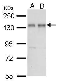
NFAT1 antibody detects NFATC2 protein by western blot analysis.
A. 30 μg Jurkat whole cell lysate/extract
B. 30 μg Raji whole cell lysate/extract
7.5% SDS-PAGE
NFAT1 antibody (GTX127932) dilution: 1:1000
The HRP-conjugated anti-rabbit IgG antibody (GTX213110-01) was used to detect the primary antibody.
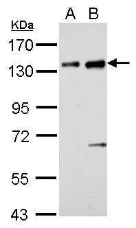
NFAT1 antibody detects NFATC2 protein by western blot analysis.
A. 30 μg BCL-1 whole cell lysate/extract
B. 30 μg Raw264.7 whole cell lysate/extract
7.5% SDS-PAGE
NFAT1 antibody (GTX127932) dilution: 1:1000
The HRP-conjugated anti-rabbit IgG antibody (GTX213110-01) was used to detect the primary antibody.
-
HostRabbit
-
ClonalityPolyclonal
-
IsotypeIgG
-
ApplicationsWB ICC/IF IP
-
ReactivityHuman, Mouse